. 2014 Jul 15;111(28):10317-22.
doi: 10.1073/pnas.1409311111. Epub 2014 Jun 30.

Genetic analysis of the Complexin trans-clamping model for cross-linking SNARE complexes in vivo

Affiliations

Genetic analysis of the Complexin trans-clamping model for cross-linking SNARE complexes in vivo

Richard W Cho et al. Proc Natl Acad Sci U S A. .

Abstract

Complexin (Cpx) is a SNARE-binding protein that regulates neurotransmission by clamping spontaneous synaptic vesicle fusion in the absence of Ca(2+) influx while promoting evoked release in response to an action potential. Previous studies indicated Cpx may cross-link multiple SNARE complexes via a trans interaction to function as a fusion clamp. During Ca(2+) influx, Cpx is predicted to undergo a conformational switch and collapse onto a single SNARE complex in a cis-binding mode to activate vesicle release. To test this model in vivo, we performed structure-function studies of the Cpx protein in Drosophila. Using genetic rescue approaches with cpx mutants that disrupt SNARE cross-linking, we find that manipulations that are predicted to block formation of the trans SNARE array disrupt the clamping function of Cpx. Unexpectedly, these same mutants rescue action potential-triggered release, indicating trans-SNARE cross-linking by Cpx is not a prerequisite for triggering evoked fusion. In contrast, mutations that impair Cpx-mediated cis-SNARE interactions that are necessary for transition from an open to closed conformation fail to rescue evoked release defects in cpx mutants, although they clamp spontaneous release normally. Our in vivo genetic manipulations support several predictions made by the Cpx cross-linking model, but unexpected results suggest additional mechanisms are likely to exist that regulate Cpx's effects on SNARE-mediated fusion. Our findings also indicate that the inhibitory and activating functions of Cpx are genetically separable, and can be mapped to distinct molecular mechanisms that differentially regulate the SNARE fusion machinery.

Keywords: exocytosis; neurotransmitter release; synapse.


Conflict of interest statement

The authors declare no conflict of interest.

Figures

Fig. 1.
Fig. 1.
The clamping and activation properties of Cpx are proposed to be mediated by a trans Cpx/SNARE array that forms between the accessory/central domains of Cpx and partially zippered prefusion SNARE complexes. (A) Structure of Cpx, and the Cpx mutants used in this study. (B) The trans Cpx/SNARE array (zigzag array) that is proposed to form between Cpx and SNARE complexes. A conformational switch in Cpx from an open (trans–SNARE) to a closed (cis–SNARE) state is proposed to mediate Ca2+-dependent triggering of vesicle fusion. The positions of mutations used in this study are marked with colors matching Fig. 1A. (C) Hydrophobic residues that mediate the interaction of the Cpx accessory helix with t-SNAREs of a partially zippered SNARE complex are conserved in both mCpx and DmCpx. Space fill model of the accessory domain of mCpx and Dm Cpx is shown. Blue and gray residues represent the interface between the Cpx accessory domain and t-SNAREs. Conserved hydrophobic residues are illustrated in gray. (D) Both mCpx and DmCpx exhibit clamping properties in in vitro cell–cell fusion assays. (E) SB mCpx exhibits clamping properties similar to WT mCpx in cell–cell fusion assays. (F) SB mCpx exhibits decreased Syt-stimulated cell–cell fusion compared with WT mCpx. Error bars are SEM, and horizontal lines indicate statistical comparisons determined using one-way ANOVA with post hoc Tukey analysis for D–F.
Fig. 2.
Fig. 2.
Mini frequency of elav-GAL4 transgenic rescued animals using WT or mutant mCpxs in the cpxSH1 mutant background. (A) Sample traces of spontaneous release events from muscle 6 of control, cpxSH1, and rescue lines expressing WT mCpx, SC mCpx, NC mCpx, SB mCpx, HB mCpx, or mCpx 51–134. (B) Summary of mean mini frequency (hertz ± SEM) for each line. Numbers in parentheses (n) represent individual NMJ recordings from indicated genotypes. Horizontal lines indicate statistical comparisons determined using ANOVA with post hoc Tukey analysis.
Fig. 3.
Fig. 3.
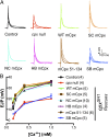
Evoked release properties of WT mCpx and individual mutant mCpx rescues. (A) Averaged traces of EJPs from control, cpxSH1, and rescued strains expressing indicated mutant mCpxs. Recordings were performed at various Ca2+ concentrations (0.15, 0.2, 0.4, and 1 mM). (B) Summary of mean EJP amplitude (millivolts ± SEM) plotted at the indicated Ca2+ concentrations. Numbers in parentheses (n) represent individual muscle recordings at the indicated [Ca2+]; data were collected from at least three independent larvae for each genotype.
Fig. 4.
Fig. 4.
SB mCpx exhibits impaired binding to postfusion, cis–DmSNAREs. (A) ∼65 µM WT mCpx was titrated into a mixture of ∼5.3-µM DmSNARE complexes. (B) ∼140 µM SB mCpx was titrated into ∼8-µM DmSNAREs. (A and B, Upper) Raw data in power vs. time during the injection after subtracting from the baseline. (Lower) Integrated heat of each injection normalized by the moles of injectant vs. the molar ratio between Cpx and SNARE in the sample cell. The solid lines represented the best fit to the black squares obtained from a nonlinear least-squares fit assuming a simple one-site chemical reaction. The results gave the thermodynamic parameters for each binding reaction, which are listed in Table 1.

References

    1. Söllner T, et al. SNAP receptors implicated in vesicle targeting and fusion. Nature. 1993;362(6418):318–324. - PubMed
    1. Weber T, et al. SNAREpins: Minimal machinery for membrane fusion. Cell. 1998;92(6):759–772. - PubMed
    1. Sutton RB, Fasshauer D, Jahn R, Brunger AT. Crystal structure of a SNARE complex involved in synaptic exocytosis at 2.4 A resolution. Nature. 1998;395(6700):347–353. - PubMed
    1. Li F, et al. Energetics and dynamics of SNAREpin folding across lipid bilayers. Nat Struct Mol Biol. 2007;14(10):890–896. - PubMed
    1. Rizo J, Rosenmund C. Synaptic vesicle fusion. Nat Struct Mol Biol. 2008;15(7):665–674. - PMC - PubMed

Publication types

LinkOut - more resources