. 2014 Jul 15;111(28):10287-92.
doi: 10.1073/pnas.1401337111. Epub 2014 Jun 30.

Engineered nanomedicine for myeloma and bone microenvironment targeting

Affiliations

Engineered nanomedicine for myeloma and bone microenvironment targeting

Archana Swami et al. Proc Natl Acad Sci U S A. .

Abstract

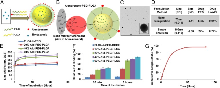
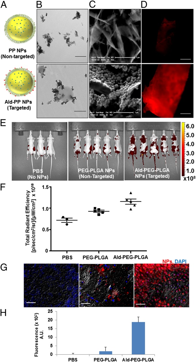
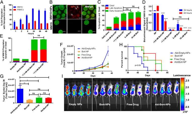
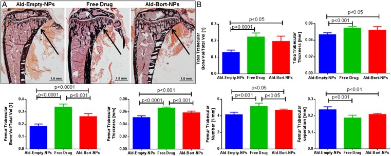
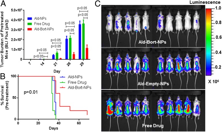
Bone is a favorable microenvironment for tumor growth and a frequent destination for metastatic cancer cells. Targeting cancers within the bone marrow remains a crucial oncologic challenge due to issues of drug availability and microenvironment-induced resistance. Herein, we engineered bone-homing polymeric nanoparticles (NPs) for spatiotemporally controlled delivery of therapeutics to bone, which diminish off-target effects and increase local drug concentrations. The NPs consist of poly(D,L-lactic-co-glycolic acid) (PLGA), polyethylene glycol (PEG), and bisphosphonate (or alendronate, a targeting ligand). The engineered NPs were formulated by blending varying ratios of the synthesized polymers: PLGA-b-PEG and alendronate-conjugated polymer PLGA-b-PEG-Ald, which ensured long circulation and targeting capabilities, respectively. The bone-binding ability of Ald-PEG-PLGA NPs was investigated by hydroxyapatite binding assays and ex vivo imaging of adherence to bone fragments. In vivo biodistribution of fluorescently labeled NPs showed higher retention, accumulation, and bone homing of targeted Ald-PEG-PLGA NPs, compared with nontargeted PEG-PLGA NPs. A library of bortezomib-loaded NPs (bone-targeted Ald-Bort-NPs and nontargeted Bort-NPs) were developed and screened for optimal physiochemical properties, drug loading, and release profiles. Ald-Bort-NPs were tested for efficacy in mouse models of multiple myeloma (MM). Results demonstrated significantly enhanced survival and decreased tumor burden in mice pretreated with Ald-Bort-NPs versus Ald-Empty-NPs (no drug) or the free drug. We also observed that bortezomib, as a pretreatment regimen, modified the bone microenvironment and enhanced bone strength and volume. Our findings suggest that NP-based anticancer therapies with bone-targeting specificity comprise a clinically relevant method of drug delivery that can inhibit tumor progression in MM.

Keywords: alendronate-PLGA-PEG; bisphosphonate; bone metastasis; targeting nanomedicine.


Conflict of interest statement

Conflict of interest statement: I.M.G. discloses her Advisory Board Membership with Novartis, Onyx, and BMS. O.C.F. discloses his financial interest in BIND Therapeutics, Selecta Biosciences, and Blend Therapeutics, three biotechnology companies developing nanoparticle technologies for medical applications. BIND, Selecta, and Blend did not support the aforementioned research, and currently these companies have no rights to any technology or intellectual property developed as part of this research.

Figures

Fig. 1.
Fig. 1.
Design, engineering, and characterization of NPs for bone targeting. (A) Schematic illustration of alendronate-conjugated PEG-PLGA (Ald-PP) NPs synthesized by blending polymers (PLGA-b-PEG-Ald and PLGA-b-PEG) in varying ratios and encapsulating the drug bortezomib. (B) Schematic representation of the mechanism of affinity of Ald-PP NPs with bone mineral (gray, bone mineral; red, Ald; green, PEG; yellow, PLGA). (C) Representative TEM image of Ald-PP NPs (single emulsion), negatively stained, imaged at 80.0 kV. (Scale bars: 500 nm; Inset, 100 nm.) (D) Physiochemical characteristics of Ald-PP NPs. (E) Size of the Ald-PP NPs (single emulsion) with varying content of polymer PLGA-b-PEG-Ald, in presence of serum, with time. (F) Quantitative evaluation of HA binding of NPs (single emulsion) with varying content of PLGA-b-PEG-Ald polymer. PLGA-b-PEG (-COOH terminated) polymeric NPs were used as control. (G) Release kinetics of encapsulated drug bortezomib from the Ald-PP NPs (single emulsion), in physiological ionic and temperature conditions.
Fig. 2.
Fig. 2.
Bone-targeting ability of Ald-PP NPs (single emulsion). (A) Schematic illustration of bone-targeted Ald-PP NPs and nontargeted PP NPs. (B) Representative TEM image of Ald-PP (Lower) NPs surface interactions with HA rods, which is not observed in case of nontargeted PP NPs (Upper). (Scale bar: 500 nm.) (C) Representative SEM image of interaction of NPs (targeted Ald-PP: Lower; nontargeted PP: Upper) with crystalized HA (scale bar: 1 µm) after incubation with NP solution and washing. (D) Representative fluorescence image of bone fragment after incubation with fluorescently labeled NP solution (targeted Ald-PP: Lower; nontargeted PP: Upper) (ex vivo), and washing. (Scale bar: 500 µm.) (E) Whole-body mice imaging (IVIS), where targeted NP (Right) clearance is compared with nontargeted NPs (Center) and PBS (Left) (24-h time point, i.p. injection). Scale represents luminescence signal from Alexa647-labeled NPs, representing NP biodistribution. (F) Total fluorescence quantified in the region of interest of IVIS images from E. (G) Representative images of bone histology in merged channels (405: DAPI; 647: NPs; and bright field) for PBS (Left), nontargeted NPs (Center), and targeted NPs (Right). (Scale bar: 100 µm.) (H) Quantification of NP homing as measured from the bone (femur and spine) histology by fluorescence intensity (average) quantification in the 647 channel in multiple sections of bone, covering entire region representatively, in different mice (n = 3).
Fig. 3.
Fig. 3.
In vitro and in vivo efficacy of NPs (single emulsion). (A) Cellular uptake of NPs during coculture with myeloma (MM1S) cells and peripheral blood mononuclear cells (PBMCs). (B) Alexa647-labeled NPs imaged in GFP+ MM1S cells using fluorescence confocal imaging. Bort-NPs induced apoptosis and death in MM1S cells (24 h) (scale bar: 5 µm) (C) as measured by Annexin-V/PI flow cytometry; and (D) bioluminescent signal quantification of GFP+Luc+ MM1S cells (24, 48 h). In C and D, cells were treated with effective bortezomib concentrations of ∼3.6 or ∼7.3 nM (Bort-NPs) or free drug (5 or 10 nM). T tests evaluating efficacy of treatments vs. NP controls at same time point show equivalent efficacy of 7.3 nM Bort-NPs and 10 nM Free Drug. (E) Annexin-V/PI flow cytometry of GFP+ MM1S cells treated with Empty-NPs, Ald-Empty-NPs, ∼3.6 nM Ald-Bort-NPs, and ∼7.3 nM Bort-NPs after 24 h. The stacked bars represent means ± SEM. (F–I) Mice injected with GFP+Luc+ MM1S cells, treated with Ald-Empty-NPs, Bort-NPs, Free Drug, and Ald-Bort-NPs twice a week, starting at day 21 after tumor cell injection (n = 7). (F) BLI flux measuring tumor burden in mice from day 21 to 38. (G) Quantification of BLI at day 38. (H) Survival data for mice treated with Bort-NPs, Ald-Bort-NPs, Free Drug, or NP controls. (I) Representative BLI images of mice at day 38 from the four groups. Scale represents luminescence signal from Luc+ MM1S cells, quantifying tumor burden.
Fig. 4.
Fig. 4.
In vivo effects of bortezomib NPs (single emulsion) on bones. Mice were pretreated for 3 wk, with Ald-Empty-NPs, Free Drug, or Ald-Bort-NPs. Static bone histomorphometry and micro-CT done on these samples show an increase in bone formation markers for the bortezomib-treated groups. (A) Representative images from static histomorphomety from each group shown by Von Kossa staining. Trabecular bone volume was increased in Free Drug and Ald-Bort-NP groups compared with that of Ald-Empty-NP group, as indicated by arrows. (B) Micro-CT analysis demonstrated significantly higher bone in Ald-Bort-NPs and Free Drug compared with that of Ald-Empty-NPs in terms of the following: tibia trabecular bone volume per total volume, tibia trabecular thickness, femur trabecular bone volume per total volume, femur trabecular thickness, femur trabecular number, and femur trabecular separation.
Fig. 5.
Fig. 5.
Pretreatment with Ald-Bort-NPs inhibits myeloma growth better than free drug. (A–C) Mice were pretreated for 3 wk with Ald-Empty-NPs, Free Drug, or Ald-Bort-NPs and then injected with GFP+Luc+ MM1S cells. (A) BLI flux from mice was significantly lower in Ald-Bort-NPs compared with that of Ald-Empty-NPs or Free Drug groups at every day of imaging. (B) Survival was also significantly increased in the Ald-Bort-NP–pretreated mice (P = 0.01). (C) Day 29 images of BLI signal from mice illustrates the reduction of tumor burden in mice pretreated with Ald-Bort-NPs (n = 10).

References

    1. Roodman GD. Pathogenesis of myeloma bone disease. Leukemia. 2009;23(3):435–441. - PubMed
    1. Reagan MR, Ghobrial IM. Multiple myeloma mesenchymal stem cells: Characterization, origin, and tumor-promoting effects. Clin Cancer Res. 2012;18(2):342–349. - PMC - PubMed
    1. Coleman RE. Metastatic bone disease: Clinical features, pathophysiology and treatment strategies. Cancer Treat Rev. 2001;27(3):165–176. - PubMed
    1. Garrett IR, et al. Selective inhibitors of the osteoblast proteasome stimulate bone formation in vivo and in vitro. J Clin Invest. 2003;111(11):1771–1782. - PMC - PubMed
    1. Ozaki S, et al. Therapy with bortezomib plus dexamethasone induces osteoblast activation in responsive patients with multiple myeloma. Int J Hematol. 2007;86(2):180–185. - PubMed

Publication types

MeSH terms