Clinical Trial
. 2014 Jul 15;111(28):10137-42.
doi: 10.1073/pnas.1405388111. Epub 2014 Jun 30.

Human endothelial colony-forming cells serve as trophic mediators for mesenchymal stem cell engraftment via paracrine signaling

Affiliations
Clinical Trial

Human endothelial colony-forming cells serve as trophic mediators for mesenchymal stem cell engraftment via paracrine signaling

Ruei-Zeng Lin et al. Proc Natl Acad Sci U S A. .

Abstract

Endothelial colony-forming cells (ECFCs) are endothelial precursors that circulate in peripheral blood. Studies have demonstrated that human ECFCs have robust vasculogenic properties. However, whether ECFCs can exert trophic functions in support of specific stem cells in vivo remains largely unknown. Here, we sought to determine whether human ECFCs can function as paracrine mediators before the establishment of blood perfusion. We used two xenograft models of human mesenchymal stem cell (MSC) transplantation and studied how the presence of ECFCs modulates MSC engraftment and regenerative capacity in vivo. Human MSCs were isolated from white adipose tissue and bone marrow aspirates and were s.c. implanted into immunodeficient mice in the presence or absence of cord blood-derived ECFCs. MSC engraftment was regulated by ECFC-derived paracrine factors via platelet-derived growth factor BB (PDGF-BB)/platelet-derived growth factor receptor (PDGFR)-β signaling. Cotransplanting ECFCs significantly enhanced MSC engraftment by reducing early apoptosis and preserving stemness-related properties of PDGFR-β(+) MSCs, including the ability to repopulate secondary grafts. MSC engraftment was negligible in the absence of ECFCs and completely impaired in the presence of Tyrphostin AG1296, an inhibitor of PDGFR kinase. Additionally, transplanted MSCs displayed fate-restricted potential in vivo, with adipose tissue-derived and bone marrow-derived MSCs contributing exclusive differentiation along adipogenic and osteogenic lineages, respectively. This work demonstrates that blood-derived ECFCs can serve as paracrine mediators and regulate the regenerative potential of MSCs via PDGF-BB/PDGFR-β signaling. Our data suggest the systematic use of ECFCs as a means to improve MSC transplantation.

Keywords: adipogenesis; angiocrine factors; osteogenesis; vasculogenesis.


Conflict of interest statement

The authors declare no conflict of interest.

Figures

Fig. 1.
Fig. 1.
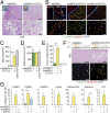
ECFCs provide trophic support to MSCs before the onset of perfusion. (A) Human ECFCs and MSCs were transplanted into nude mice. Rhodamine-conjugated UEA-1 was i.v.-injected into implant-bearing mice 10 min before harvesting the implants. Retrieved cells were identified by flow cytometry as follows: nonhematopoietic cells (mCD45 cells; R1 gate); human MSCs (mCD45/hCD90+/hCD31 cells; R2 gate); total human ECFCs (mCD45/hCD90/hCD31+ cells; R3 gate); and perfused (UEA-1+) ECFCs (mCD45/hCD90/hCD31+/UEA-1+ cells; R4 gate). (B) Cytometric quantification of perfused human ECFCs in explanted grafts. (C) Projections of whole-mount fluorescent staining of explanted grafts. ECFCs were identified by expression of hCD31 (green) and perfused ECFCs by UEA-1 (red). (Scale bars, 100 μm.) (D) MSCs were implanted into GFP-SCID mice with or without ECFCs. Viable (PI/annexin-V) and apoptotic (annexin-V+) MSCs were identified at day 2 by flow cytometry. (E) Percentage of apoptotic MSCs at day 2 in grafts with or without ECFCs and ECFC-CM. (F) Number of viable MSCs at day 7 in grafts with or without ECFCs. Bars represent mean ± SEM. Mice per group: n = 4. *P < 0.05, **P < 0.01, and ***P < 0.001 compared with grafts with MSCs alone.
Fig. 2.
Fig. 2.
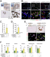
Loss of MSC properties in the absence of early angiocrine support. (A) MSCs were transplanted into GFP-SCID mice with or without ECFCs and then retrieved at day 7 and selected as hCD90+ cells (n = 3 per group). (B) Enhanced CFU-F activity in MSCs from ECFC-containing grafts (5 × 10² MSCs plated per group). (C) Quantitative CFU-F activity of retrieved MSCs. (D) Ex vivo adipogenic and osteogenic differentiation of retrieved MSCs at clonal density (n = 16 clones per group). (E) Percentage of MSC clones with single and double differentiation potential after retrieval from grafts. Bars represent mean ± SEM. *P < 0.05 compared with grafts with MSCs alone.
Fig. 3.
Fig. 3.
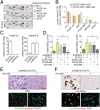
Irreversible loss of watMSC adipogenic potential in the absence of initial angiocrine support. (A) MSCs were transplanted into nude mice with or without ECFCs for 28 d, and H&E staining of grafts was performed. (Insets) Macroscopic explants. (B) Immunohistochemical analysis of adipocytes in explanted grafts. Adipocytes express perilipin-A (red). Human MSC-derived cells express h-vimentin (green). Yellow asterisks: human adipocytes. White asterisks: murine adipocytes. (C) Quantification of adipocyte density. (D) Adipocyte size in explants. (E) Percentage of human adipocytes (perilipin-A+/h-vimentin+). (F) watMSCs were transplanted into primary GFP-SCID mice for 7 d and then retrieved and transplanted with ECFCs into secondary nude mice for 28 d. H&E and immunofluorescence staining was used for secondary grafts. Yellow asterisks: human adipocytes. White asterisks: murine adipocytes. (G) mRNA expression of adipogenic factors in explanted grafts. All primers were human-specific (SI Appendix, Table S1). Data are normalized to human β-actin (ACTB). [Scale bars: 200 μm (A, B, and F, Upper); 50 μm (A, B, and F, Lower); and 5 mm (A, Insets).] Bars represent mean ± SEM (n = 4 mice per group). *P < 0.05. ***P < 0.001 compared with grafts with MSCs alone.
Fig. 4.
Fig. 4.
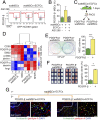
Irreversible loss of bmMSC osteogenic potential in the absence of initial angiocrine support. (A) MSCs were implanted into nude mice with or without ECFCs and BMP-2 for 28 d. von Kossa staining was used for the grafts. Top panels are montages of contiguous pictures capturing the entire cross-section of the explants, with dashed black lines delineating the border of each mounted picture. (Insets) Macroscopic views of the explants. (B) Immunohistochemical analysis of osteogenic differentiation. Osteoblasts express osterix (red). Human MSC-derived cells express h-vimentin (green). Yellow arrowheads: human osteoblasts. White arrowheads: murine osteoblasts. (C) Quantification of tissue mineralization. (D) Percentage of human osteoblasts (osterix+/h-vimentin+). (E) bmMSCs were transplanted into primary GFP-SCID mice for 7 d and then retrieved and transplanted with ECFCs into secondary nude mice for 28 d. von Kossa and immunofluorescence staining was used for BMP-2–stimulated secondary grafts. Yellow arrowheads: human osteoblasts. White arrowheads: murine osteoblasts. (F) mRNA expression of osteogenic factors. All primers were human-specific (SI Appendix, Table S1). Data are normalized to human β-actin (ACTB). [Scale bars: 1 mm (A, Upper); 200 μm (A, Lower; E, Upper); 3 mm (A, Insets); 50 μm (B, Upper); and 10 μm (B and E, Lower).] Bars represent mean ± SEM (n = 4 mice per group). *P < 0.05. **P < 0.01 compared with grafts with MSCs alone.
Fig. 5.
Fig. 5.
ECFCs support MSC engraftment via paracrine secretion of PDGF-BB. (A) Protein arrays of conditioned media from ECFCs, MSCs, and MSCs+ECFCs (1:1 ratio). Color-lined boxes indicate ECFC-secreted angiocrine factors. (B) Quantification of angiocrine factors: (i) conditioned medium from ECFC-MSC coculture [(ECFC-MSC)-CM]; (ii) A 1:1 mixture of conditioned media from monocultures of ECFCs and MSCs (ECFC-CM+MSC-CM). (C) mRNA expression of PDGF-B and PDGFR-β in ECFCs, watMSCs, and bmMSCs. Data normalized to ribosomal 18S rRNA. (D) Flow cytometry quantification of viable MSCs at day 2 in grafts with or without ECFCs, ECFC-CM, PDGF-BB, and PDGFR inhibitor (AG1296). (E) MSCs were transplanted into nude mice with ECFCs with or without AG1296. H&E and immunofluorescence staining were performed at day 28. Human adipocytes: perilipin-A+/h-vimentin+ cells (yellow asterisks). (F) MSCs were transplanted into nude mice with ECFCs, BMP-2, and with or without AG1296. von Kossa and immunofluorescence staining were performed at day 28. Human osteoblasts: osterix+/h-vimentin+ cells (yellow arrowheads). [Scale bars: 200 μm (E and F, Upper) and 50 μm (E and F, Lower).] Bars represent mean ± SEM (n = 4 mice per group). *P < 0.05, **P < 0.01, and ***P < 0.001 compared with grafts with MSCs alone. P < 0.01 compared with grafts with MSCs+ECFCs.
Fig. 6.
Fig. 6.
PDGFR-β expression in transplanted MSCs coincides with progenitor cell function. (A) Flow cytometric detection of PDGFR-β+ watMSCs in grafts with or without ECFCs and PDGFR inhibitor (AG1296). (B) Percentage of PDGFR-β+ watMSCs at day 7. (C) watMSCs were transplanted into GFP-SCID mice with ECFCs, retrieved at day 7, and immediately sorted into PDGFR-β+ and PDGFR-β cells. (D) Heat map and hierarchical clustering of sorted watMSCs. mRNA data are normalized to ribosomal 18S rRNA. Human SMCs and NHDFs served as controls. (E) CFU-F activity of sorted watMSCs. (F) Ex vivo adipogenic differentiation potential of sorted watMSCs. (G) watMSCs were transplanted into primary mice with ECFCs. Retrieved watMSCs (day 7) were sorted into PDGFR-β+ and PDGFR-β cells and separately transplanted with ECFCs into secondary mice for 28 d. Immunofluorescence staining of secondary grafts was performed. Yellow asterisks: human adipocytes (perilipin-A+/h-vimentin+). White asterisks: murine adipocytes (perilipin-A+/h-vimentin). [Scale bars: 200 μm (G, Left panels) and 50 μm (G, Right panels).] Bars represent mean ± SEM. Mice per group: n = 4 (B), n = 3 (DG). Clones per group: n = 16 (F). *P < 0.05; ***P < 0.001.

References

    1. Lin Y, Weisdorf DJ, Solovey A, Hebbel RP. Origins of circulating endothelial cells and endothelial outgrowth from blood. J Clin Invest. 2000;105(1):71–77. - PMC - PubMed
    1. Ingram DA, et al. Identification of a novel hierarchy of endothelial progenitor cells using human peripheral and umbilical cord blood. Blood. 2004;104(9):2752–2760. - PubMed
    1. Melero-Martin JM, et al. In vivo vasculogenic potential of human blood-derived endothelial progenitor cells. Blood. 2007;109(11):4761–4768. - PubMed
    1. Kaushal S, et al. Functional small-diameter neovessels created using endothelial progenitor cells expanded ex vivo. Nat Med. 2001;7(9):1035–1040. - PMC - PubMed
    1. Melero-Martin JM, et al. Engineering robust and functional vascular networks in vivo with human adult and cord blood-derived progenitor cells. Circ Res. 2008;103(2):194–202. - PMC - PubMed

Publication types

MeSH terms

Substances