. 2014 Jul 15;111(28):E2851-7.
doi: 10.1073/pnas.1407777111. Epub 2014 Jun 30.

Two miRNA clusters, miR-34b/c and miR-449, are essential for normal brain development, motile ciliogenesis, and spermatogenesis

Affiliations

Two miRNA clusters, miR-34b/c and miR-449, are essential for normal brain development, motile ciliogenesis, and spermatogenesis

Jingwen Wu et al. Proc Natl Acad Sci U S A. .

Abstract

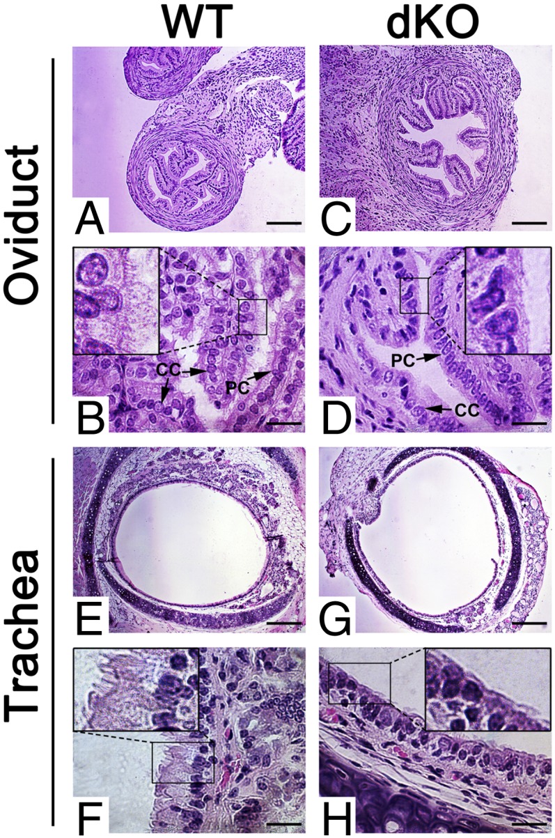
Ablation of a single miRNA gene rarely leads to a discernable developmental phenotype in mice, in some cases because of compensatory effects by other functionally related miRNAs. Here, we report that simultaneous inactivation of two functionally related miRNA clusters (miR-34b/c and miR-449) encoding five miRNAs (miR-34b, miR-34c, miR-449a, miR-449b, and miR-449c) led to sexually dimorphic, partial perinatal lethality, growth retardation, and infertility. These developmental defects correlated with the dysregulation of ∼ 240 target genes, which are mainly involved in three major cellular functions, including cell-fate control, brain development and microtubule dynamics. Our data demonstrate an essential role of a miRNA family in brain development, motile ciliogenesis, and spermatogenesis.

Keywords: airway obstruction; egg transport; forebrain; oviduct.


Conflict of interest statement

The authors declare no conflict of interest.

Figures

Fig. 1.
Fig. 1.
Expression profiles of the five miRNAs (miR-34b, miR-34c, miR-449a, miR-449b, and miR-449c) encoded by the miR-34b/c and miR-449 clusters in multiple organs of WT and KO mice. (A) Relative expression levels of the five miRNAs in 11 organs of WT mice. Expression levels of the five miRNAs were first determined using TaqMan-based qPCR analyses. Relative levels are represented by fold changes, which were calculated by designating the levels in lung as 1. Undetectable Ct values were considered no expression. (B–E) Fold changes in expression levels of the five miRNAs in single-KO (miR-34b/c−/− or miR-449−/−) and double-KO (miR-34−/−; miR-449−/−) mice compared with the WT controls. Expression levels of the five miRNAs were first determined using TaqMan-based qPCR analyses, and fold changes were then calculated by designating the WT levels as 1. Four organs expressing the five miRNAs (A), including brain (B), testis (C), ovary (D), and lung (E), were analyzed. In miRNA qPCR analyses, U6 was used as a loading control, and values were calculated based on the relative quantification (ΔΔCt) method. All qPCR assays were performed in biological triplicates.
Fig. 2.
Fig. 2.
Partial perinatal lethality and growth retardation in miR-34b/c and miR-449 dKO mice. (A) Lethality rate of double heterozygous and “triple negative” (miR-34b/c−/−; miR-449+/− or miR-34b/c+/−; miR-449−/−) littermate control (con) and miR-34b/cmiR-449 dKO male (M) and female (F) pups around P7. P values < 0.01 are statistically significant. Data were based on 383 pups produced by triple-negative breeding pairs. (B) Growth curves showing the changes in body weight of double-heterozygous and triple-negative littermate control (con) and dKO male (M) and female (F) mice over the first 20 d of postnatal development (P1–P20). The number of pups (n) measured is indicated. Mean values are presented. Original mean values, SD, and P values are presented in SI Appendix, Table S1. (C) Body weight of control (con) and dKO male and dKO female mice at age 4 and 8 wk. Data are presented as mean ± SEM (n = 6). P values < 0.01 are statistically significant.
Fig. 3.
Fig. 3.
Defects in basal forebrain structures in dKO mice. (A and B) Schematic diagram of the male basal forebrain at E18.5 showing differences in the area of the CPu and the OT between control and dKO brains. The dKO CPu area is ∼89.7% of the control, and the dKO OT is ∼55.8% of the control. (C and D) DAPI labeling of sagittal sections of the CPu at E18.5 showing that the dKO brains (n = 4) display narrow CPu regions and smaller OTs compared with control (n = 4). (Scale bars: 200 μm.) (E–H) High-magnification images of the control and dKO CPu and OT regions labeled with DAPI (E–H), βIII tubulin (E′–H′), and Nissl (E′′–H′′) in sagittal sections showing reduced size in the dKO brains compared with control. (Scale bars: 200 μm.) Pir, piriform cortex; Acb, accumbens nucleus.
Fig. 4.
Fig. 4.
Disrupted spermatogenesis and oligoasthenoteratozoospermia in male dKO mice, and oviduct defects in female dKO mice. (A–D) Testicular histology of 10-wk-old WT mice (A and B) and dKO mice (C and D). (Scale bar: 20 μm.) (E) Sperm counts of WT and dKO male mice. Data are presented as mean ± SEM (n = 6). (F) Deformed sperm in dKO male mice. (Scale bar: 20 μm.) (G and H) Pie charts showing the proportional distribution of normal and deformed sperm in WT (G) and dKO (H) male mice. (I) Normal ovarian histology of dKO female mice at P60. (Scale bar: 50 μm.) (J) Number of MII oocytes retrieved from the oviducts of adult WT and dKO female mice after superovulation. Data are presented as mean ± SEM (n = 6). (K) Number of germinal vesicle (GV)-stage oocytes retrieved from the PMSG-primed ovaries of adult WT and adult dKO female mice. Data are presented as mean ± SEM (n = 6). (L) Maturation rate of WT and dKO oocytes from GV to meiosis I (MI) and meiosis II (MII) stages during a 20-h culture in vitro. Data are presented as mean ± SEM (n = 6).
Fig. 5.
Fig. 5.
Deficiency in motile ciliogenesis in dKO mice. (A–D) Although the WT oviduct epithelia consist of both Peg cells (PC) and ciliated cells (CC) with abundant cilia pointing to the lumen (A and B), CCs and cilia are rarely seen despite the presence of PCs in the epithelia of dKO oviducts (C and D). (Insets) Digitally enlarged, framed areas in B and D. (Scale bars: 200 μm in A and C, 20 μm in B and D.) Five WT and dKO mice were analyzed, and representative images are shown. (E–H) The pseudostratified columnar epithelia of the WT trachea contain abundant cilia pointing to the lumen (E and F), whereas cilia are largely lacking in the epithelia of the dKO trachea (G and H). (Insets) Digitally enlarged and framed areas in F and H. (Scale bars: 200 μm in E and G, 20 μm in F and H). Five WT and dKO mice were analyzed, and representative images are shown.
Fig. 6.
Fig. 6.
GO term enrichment analyses. Analyses of dysregulated genes in dKO brains and testes reveal that the five miRNAs control 239 target mRNAs involved in three major cellular functions, including brain development, cell fate control, and cytoskeleton dynamics. Disrupted brain development, motile ciliogenesis, and spermatogenesis in dKO mice result from both the primary and secondary effects of dysregulated target genes, owing to ablation of the five miRNAs.

References

    1. Bartel DP. MicroRNAs: Target recognition and regulatory functions. Cell. 2009;136(2):215–233. - PMC - PubMed
    1. Bartel DP. MicroRNAs: Genomics, biogenesis, mechanism, and function. Cell. 2004;116(2):281–297. - PubMed
    1. Loh YH, Yi SV, Streelman JT. Evolution of microRNAs and the diversification of species. Genome Biol Evol. 2011;3:55–65. - PMC - PubMed
    1. Janga SC, Vallabhaneni S. MicroRNAs as post-transcriptional machines and their interplay with cellular networks. Adv Exp Med Biol. 2011;722:59–74. - PubMed
    1. Peters J, Robson JE. Imprinted noncoding RNAs. Mamm Genome. 2008;19(7-8):493–502. - PubMed

Publication types