

Molecular and nonmolecular diagnostic methods for invasive fungal infections
- PMID: 24982319
- PMCID: PMC4135902
- DOI: 10.1128/CMR.00091-13
Molecular and nonmolecular diagnostic methods for invasive fungal infections
Abstract
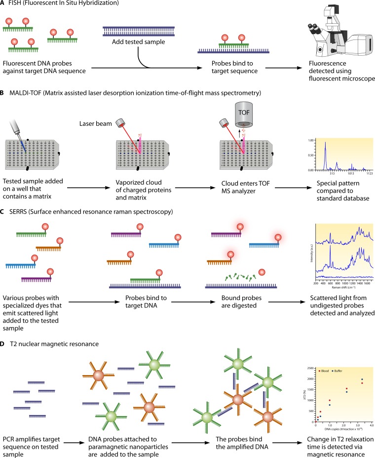
Invasive fungal infections constitute a serious threat to an ever-growing population of immunocompromised individuals and other individuals at risk. Traditional diagnostic methods, such as histopathology and culture, which are still considered the gold standards, have low sensitivity, which underscores the need for the development of new means of detecting fungal infectious agents. Indeed, novel serologic and molecular techniques have been developed and are currently under clinical evaluation. Tests like the galactomannan antigen test for aspergillosis and the β-glucan test for invasive Candida spp. and molds, as well as other antigen and antibody tests, for Cryptococcus spp., Pneumocystis spp., and dimorphic fungi, have already been established as important diagnostic approaches and are implemented in routine clinical practice. On the other hand, PCR and other molecular approaches, such as matrix-assisted laser desorption ionization (MALDI) and fluorescence in situ hybridization (FISH), have proved promising in clinical trials but still need to undergo standardization before their clinical use can become widespread. The purpose of this review is to highlight the different diagnostic approaches that are currently utilized or under development for invasive fungal infections and to identify their performance characteristics and the challenges associated with their use.
Copyright © 2014, American Society for Microbiology. All Rights Reserved.
Figures


References
-
- Pappas PG, Alexander BD, Andes DR, Hadley S, Kauffman CA, Freifeld A, Anaissie EJ, Brumble LM, Herwaldt L, Ito J, Kontoyiannis DP, Lyon GM, Marr KA, Morrison VA, Park BJ, Patterson TF, Perl TM, Oster RA, Schuster MG, Walker R, Walsh TJ, Wannemuehler KA, Chiller TM. 2010. Invasive fungal infections among organ transplant recipients: results of the Transplant-Associated Infection Surveillance Network (TRANSNET). Clin. Infect. Dis. 50:1101–1111. 10.1086/651262 - DOI - PubMed
-
- Nguyen MH, Wissel MC, Shields RK, Salomoni MA, Hao B, Press EG, Shields RM, Cheng S, Mitsani D, Vadnerkar A, Silveira FP, Kleiboeker SB, Clancy CJ. 2012. Performance of Candida real-time polymerase chain reaction, beta-d-glucan assay, and blood cultures in the diagnosis of invasive candidiasis. Clin. Infect. Dis. 54:1240–1248. 10.1093/cid/cis200 - DOI - PubMed
-
- Baddley JW, Andes DR, Marr KA, Kontoyiannis DP, Alexander BD, Kauffman CA, Oster RA, Anaissie EJ, Walsh TJ, Schuster MG, Wingard JR, Patterson TF, Ito JI, Williams OD, Chiller T, Pappas PG. 2010. Factors associated with mortality in transplant patients with invasive aspergillosis. Clin. Infect. Dis. 50:1559–1567. 10.1086/652768 - DOI - PMC - PubMed
Publication types
MeSH terms
LinkOut - more resources
Full Text Sources
Other Literature Sources
Medical
Miscellaneous
