. 2014 Sep 1;127(Pt 17):3805-16.
doi: 10.1242/jcs.150458. Epub 2014 Jun 30.

Conformational remodeling of the fibronectin matrix selectively regulates VEGF signaling

Affiliations

Conformational remodeling of the fibronectin matrix selectively regulates VEGF signaling

Anthony Ambesi et al. J Cell Sci. .

Abstract

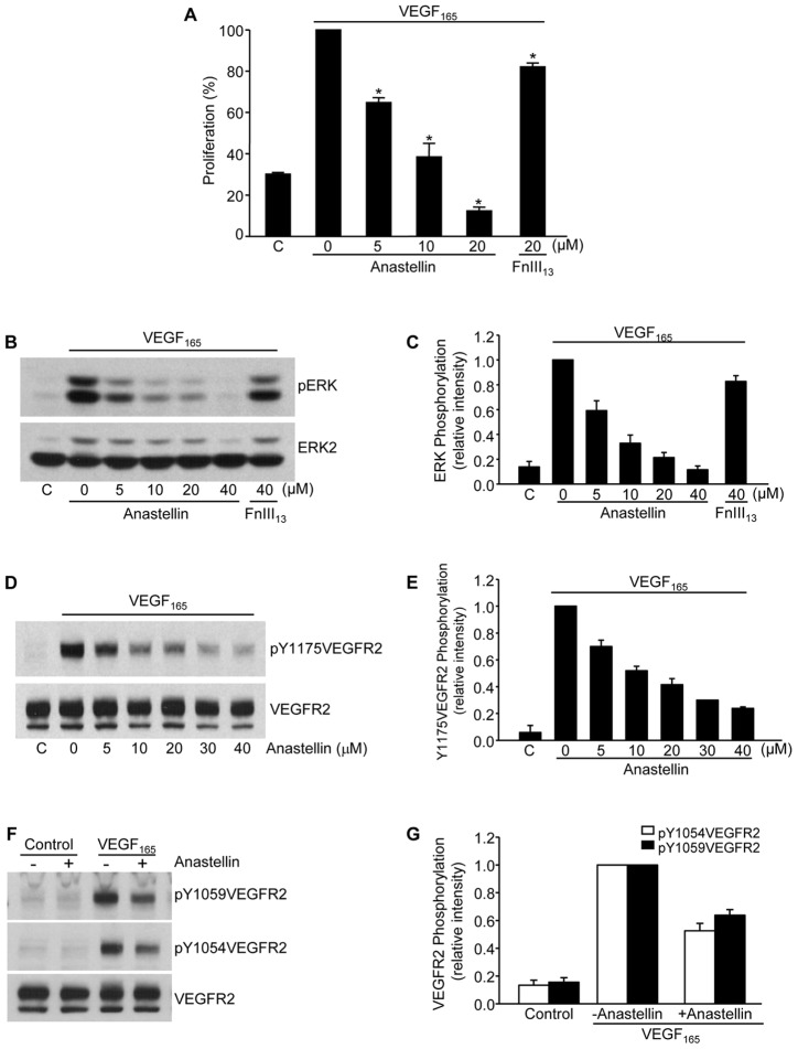
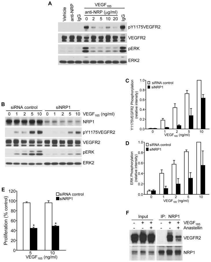
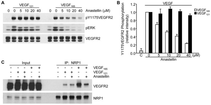
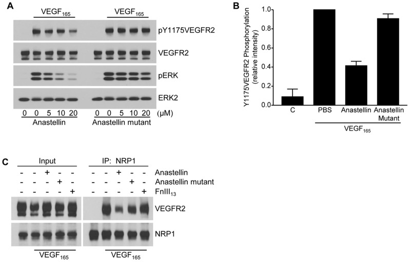
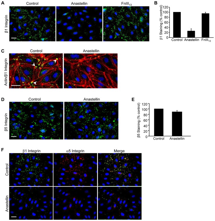
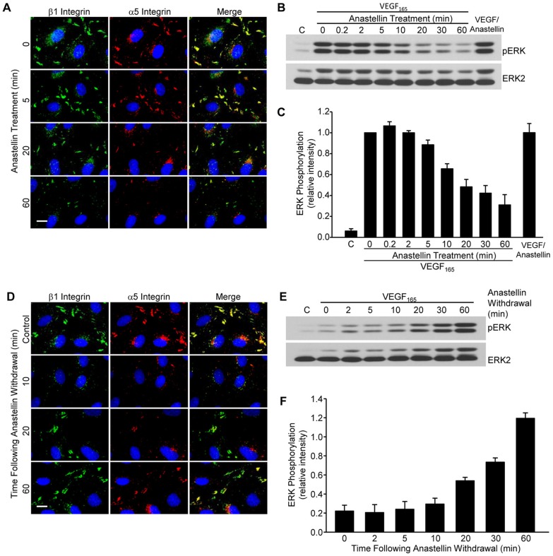
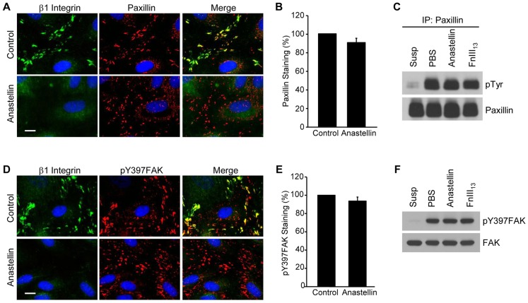
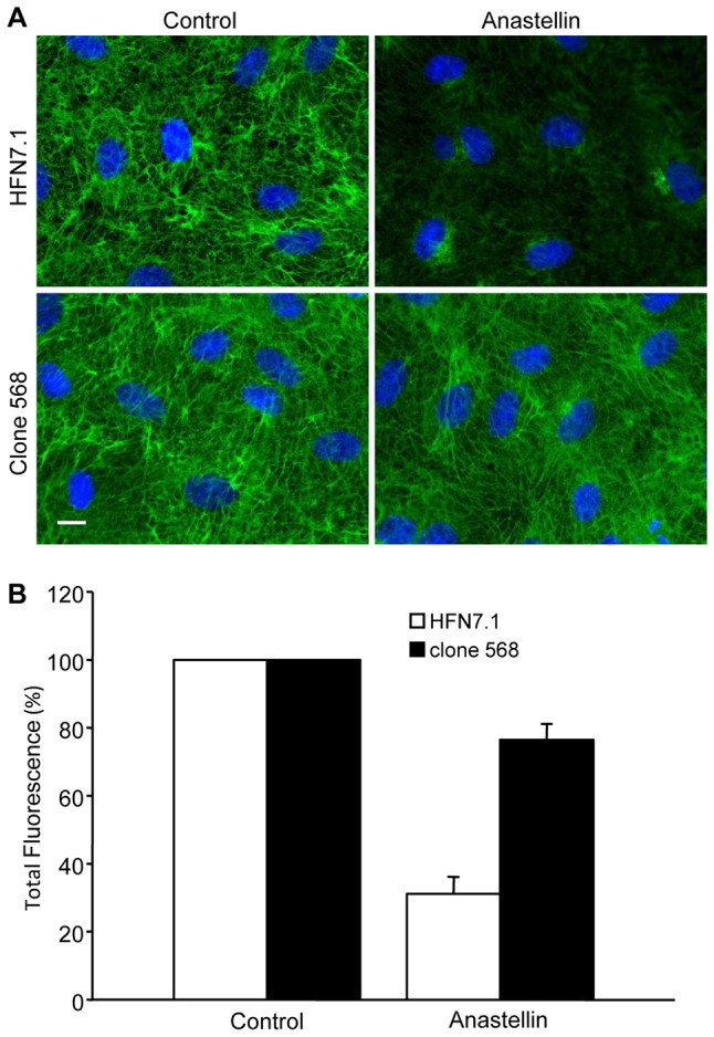
The fibronectin matrix plays a crucial role in the regulation of angiogenesis during development, tissue repair and pathogenesis. Previous work has identified a fibronectin-derived homophilic binding peptide, anastellin, as an effective inhibitor of angiogenesis; however, its mechanism of action is not well understood. In the present study, we demonstrate that anastellin selectively inhibits microvessel cell signaling in response to the VEGF165 isoform, but not VEGF121, by preventing the assembly of the complex containing the VEGF receptor and neuropilin-1. Anastellin treatment resulted in the inactivation of α5β1 integrins but was not accompanied by a change in either adhesion complexes or adhesion-based signaling. Integrin inactivation was associated with a masking of the fibronectin synergy site within the extracellular matrix (ECM), indicating that α5β1 inactivation resulted from a decrease in available ligand. These data demonstrate that anastellin influences the microvessel cell response to growth factors by controlling the repertoire of ligated integrins and point to anastellin as an effective regulator of fibronectin matrix organization. These studies further suggest that homophilic fibronectin binding peptides might have novel applications in the field of tissue regeneration as tools to regulate neovascularization.

Keywords: Anastellin; Angiogenesis; Extracellular matrix; Fibronectin; Integrin; Neuropilin; VEGF.


Figures

Fig. 1.
Fig. 1.
Anastellin inhibits VEGF-dependent microvessel endothelial cell proliferation and receptor activation. (A) Human dermal microvessel cells were cultured for 3 days in medium supplemented with or without (‘C’) 10 ng/ml VEGF165 in the presence of increasing concentrations of anastellin. FnIII13 served as a negative control. Cells were fixed and proliferation was determined by Toluidine Blue staining. Anastellin significantly inhibited proliferation in response to VEGF165 at all doses. Data show the mean±s.e.m.; *P<0.001. (B,D,F) Microvessel cells were starved overnight in serum-free medium containing 0.5% BSA and pretreated for 60 minutes with increasing concentrations of anastellin prior to stimulation with 10 ng/ml VEGF165 for 6 minutes. Cell lysates were analyzed by western blotting for phospho-ERK1/2 and VEGFR2 phosphorylation. Total ERK2 and VEGFR2 staining served as loading controls. (C,E,G) Western blots from the experiments shown in panels B,D and F were quantified using ImageJ software. Data show the mean±s.e.m. (three independent experiments).
Fig. 2.
Fig. 2.
Anastellin disrupts VEGFR2–NRP1 complex assembly and signaling. (A) Serum-starved microvessel cell monolayers were pretreated for 2 hours with increasing concentrations of an NRP1-function-blocking antibody and then stimulated with 5 ng/ml VEGF165 for 6 minutes. Cell lysates were analyzed by western blotting for phosphorylation of VEGFR2 and ERK. (B) Microvessel cells treated with either siRNA against NRP1 or control siRNA were stimulated with increasing concentrations of VEGF165 for 6 minutes and analyzed for the phosphorylation of VEGFR2 and ERK by western blotting. Total VEGFR2 and ERK2 served as loading controls. (C,D) Blots from B were quantified using ImageJ software. Data show the mean±s.e.m. (three independent experiments). (E) Microvessel cells were grown for 3 days in basal medium containing the indicated amount of VEGF165. Cells were fixed and cell counts were obtained by Toluidine Blue staining. Knockdown of NRP1 significantly inhibited cell growth. Data show the mean±s.e.m. (three independent experiments); *P<0.001. (F) Microvessel cells were treated for 60 minutes with 20 µM anastellin prior to stimulation with 10 ng/ml VEGF165 for 15 minutes. Cell monolayers were washed, and NRP1 was immunoprecipitated (IP) from cell lysates. Immunoprecipitates were analyzed by western blotting for co-precipitation of VEGFR2. Input amounts of VEGFR2 and NRP1 present in lysates prior to immunoprecipitation are shown on the left.
Fig. 3.
Fig. 3.
Anastellin has no effect on VEGF121-induced VEGFR2 activation and signaling. (A) Serum-starved microvessel cell monolayers were pretreated for 60 minutes with increasing concentrations of anastellin, followed by stimulation with either 10 ng/ml VEGF121 or VEGF165 for 6 minutes. Cell lysates were analyzed by western blotting for changes in the phosphorylation of VEGFR2 and ERK. Total levels of VEGFR2 served as a loading control. (B) Blots were quantified using ImageJ software. Data show the mean±s.e.m. (three independent experiments). ‘C’, untreated control. (C) Microvessel cell monolayers were pretreated with 20 µM anastellin for 60 minutes prior to stimulation with 10 ng/ml VEGF165 or VEGF121 for 15 minutes. NRP1 was immunoprecipitated (IP) from all lysates, and precipitates were analyzed by western blotting for the presence of VEGFR2. Input amounts of VEGFR2 and NRP1 present in lysates prior to immunoprecipitation are shown on the left.
Fig. 4.
Fig. 4.
Anastellin inhibition of VEGF165 signaling requires fibronectin matrix remodeling. (A) Serum-starved microvessel cell monolayers were pretreated with increasing concentrations of anastellin or the anastellin mutant for 60 minutes prior to stimulation with 10 ng/ml VEGF165 for 6 minutes. Cell lysates were analyzed by western blotting for phosphorylated ERK and VEGFR2. Total VEGFR2 and ERK2 served as loading controls. (B) VEGF-dependent phosphorylation of Y1175VEGFR2 in the presence of 20 µm anastellin or anastellin mutant was quantified by scanning blots from three separate experiments. ‘C’, untreated control. Data show the mean±s.e.m. (C) Microvessel cells were treated with 20 µM anastellin, anastellin mutant or the control fibronectin module FnIII13 for 60 minutes prior to stimulation with 10 ng/ml VEGF165 for 15 minutes. NRP1 was immunoprecipitated (IP) from cell lysates. Immunoprecipitates were analyzed by western blotting for co-precipitation of VEGFR2. Input amounts of VEGFR2 and NRP1 present in lysates prior to immunoprecipitation are shown on the left.
Fig. 5.
Fig. 5.
Anastellin causes inactivation of β1 integrin. Microvessel cells were treated with vehicle (control), 20 µM anastellin or the control fibronectin module FnIII13 for 60 minutes. (A) Cells were washed, fixed and stained for active β1 integrin using the monoclonal antibody 9EG7. Nuclei were counterstained with Hoechst 33258. (B) NIH ImageJ software was used to quantify the number of β1-containing focal adhesions obtained from at least five fields of view. Data are shown normalized to control values. Data averaged over five independent experiments are represented as the mean±s.e.m.; *P<0.001. (C) Cells were stained for both actin (red) and active β1 integrin (green) using phalloidin and the 9EG7 antibody, respectively. Control cells show β1 integrin localized at actin termini in focal adhesions (arrowheads). (D,E) Microvessel cells were stained with a monoclonal antibody directed against integrin β5 and quantified as in B. (F) Microvessel cells were dual stained for β1 (9EG7, green) and α5 (AB1949, red) integrin subunits. Merged images are shown on the right. Scale bars: 25 µm.
Fig. 6.
Fig. 6.
Inactivation of α5β1 integrin and VEGFR2 by anastellin is both time dependent and reversible. (A) Microvessel cells were treated with 20 µM anastellin for the times indicated, washed, fixed and stained for β1 (9EG7, green) and α5 (AB1949, red) integrins. Nuclei were counterstained with Hoechst 33258. (B) Serum-starved microvessel cells were pretreated with 20 µM anastellin for increasing amounts of time up to 60 minutes prior to stimulation with 10 ng/ml VEGF165 for 6 minutes. Alternatively, VEGF165 and anastellin (VEGF/Anastellin) were mixed (10 ng/ml VEGF165, 20 µM anastellin) and incubated for 30 minutes prior to addition to cell monolayers for 6 minutes. ‘C’, vehicle (PBS) control. Phosphorylation of ERK was examined by western blotting. Blots were stripped and reprobed for ERK2, which served as a loading control. (C) ERK phosphorylation was quantified from three independent experiments using ImageJ software and was normalized to pERK levels obtained in the absence of anastellin. Data show the mean±s.e.m. (D) Microvessel cells were treated with 20 µM anastellin for 60 minutes and were incubated with serum-free medium (no anastellin) for the indicated times. Cells were then washed, fixed and dual stained for integrin β1 (9EG7, green) and α5 (AB1949, red) subunits. Nuclei were counterstained with Hoescht 33258. Control cells received no anastellin treatment. Scale bars: 10 µm. (E) Microvessel cells were pretreated with 20 µM anastellin for 60 minutes. The medium was replaced with serum-free medium. At the indicated times, cells were stimulated with 10 ng/ml VEGF165 for 6 minutes, and lysates were analyzed by western blotting for phosphorylated ERK. Blots were stripped and reprobed for ERK2, which served as loading control. (F) ERK phosphorylation was quantified from three independent experiments using ImageJ software and was normalized to VEGF-stimulated levels of pERK observed in the absence of anastellin (data not shown). Data show the mean±s.e.m.
Fig. 7.
Fig. 7.
Anastellin does not decrease the number of focal adhesion complexes. (A) Microvessel cell monolayers were treated with 20 µM anastellin for 60 minutes and stained for both active β1 integrin (green) and paxillin (red). (B) Microvessel cells were treated as in A, and paxillin-containing contacts were quantified by ImageJ software as described for Fig. 5. Data show the mean±s.e.m. (six fields of view, four independent experiments). (C) Paxillin was immunoprecipitated (IP) from microvessel cell lysates prepared from cells treated with 20 µM anastellin or the control fibronectin module, FnIII13. Lysates prepared from cells kept in suspension (Susp) served as a control for the loss of adhesion, whereas those prepared from cells treated with PBS served as adherent untreated control. Immunoprecipitates were analyzed by western blotting for phosphotyrosine (pTyr) using the monoclonal antibody 4G10. Blots were stripped and reprobed for paxillin, which served as the loading control. (D) Cells were treated as described in A and stained with antibody against phosphorylated FAK (pTyr397FAK, red). Scale bars: 10 µm. (E) pTyr397FAK-containing focal adhesions were quantified by ImageJ software. Data show the mean±s.e.m. (six fields of view, three independent experiments). (F) Lysates from cells treated as described in C were probed for pTyr397FAK by western blotting. Blots were stripped and reprobed for total FAK, which served as loading control.
Fig. 8.
Fig. 8.
Anastellin decreases the availability of the synergy site epitope in matrix fibronectin. (A) Microvessel cell monolayers were treated with 5 µM anastellin for 60 minutes, fixed, permeabilized and stained using a fibronectin-blocking antibody (HFN7.1) against the synergy site in the FnIII9 module or an antibody (clone 568) that binds to the FnIII8 module. Scale bar: 10 µm. (B) Total fluorescence was quantified using NIH ImageJ software. Data show the mean±s.e.m. (at least six fields of view, two independent experiments).

References

    1. Ambesi A., Klein R. M., Pumiglia K. M., McKeown-Longo P. J. (2005). Anastellin, a fragment of the first type III repeat of fibronectin, inhibits extracellular signal-regulated kinase and causes G(1) arrest in human microvessel endothelial cells. Cancer Res. 65, 148–156 - PubMed
    1. Aota S., Nomizu M., Yamada K. M. (1994). The short amino acid sequence Pro-His-Ser-Arg-Asn in human fibronectin enhances cell-adhesive function. J. Biol. Chem. 269, 24756–24761 - PubMed
    1. Argraves W. S., Drake C. J. (2005). Genes critical to vasculogenesis as defined by systematic analysis of vascular defects in knockout mice. Anat. Rec. A Discov. Mol. Cell. Evol. Biol. 286A, 875–884 10.1002/ar.a.20232 - DOI - PubMed
    1. Astrof S., Hynes R. O. (2009). Fibronectins in vascular morphogenesis. Angiogenesis 12, 165–175 10.1007/s10456-009-9136-6 - DOI - PMC - PubMed
    1. Ballmer-Hofer K., Andersson A. E., Ratcliffe L. E., Berger P. (2011). Neuropilin-1 promotes VEGFR-2 trafficking through Rab11 vesicles thereby specifying signal output. Blood 118, 816–826 10.1182/blood-2011-01-328773 - DOI - PubMed

Publication types

MeSH terms

LinkOut - more resources