Case Reports
. 2014 Jul;27(3):253-6.
doi: 10.1080/08998280.2014.11929129.

Clinical and morphologic findings in disseminated Scedosporium apiospermum infections in immunocompromised patients

Affiliations
Case Reports

Clinical and morphologic findings in disseminated Scedosporium apiospermum infections in immunocompromised patients

Molly M Campa-Thompson et al. Proc (Bayl Univ Med Cent). 2014 Jul.

Abstract

Scedosporium apiospermum is a ubiquitous, saprophytic, filamentous mold that may cause localized, subcutaneous infections in immunocompetent hosts, but disseminated infection in severely immunocompromised patients. This mold is often highly resistant to multiple commonly used antifungal drugs. Even with treatment, there is a high mortality rate. We present two patients with fatal disseminated S. apiospermum infections after bone marrow and lung transplantation. This infection can be rapidly fatal, and survival may be improved by early recognition.


Figures

Figure 1.
Figure 1.
Radiographic images from Case 1. (a) An initial contrast-enhanced CT demonstrated a single 2.6 cm intraparenchymal ring-enhancing lesion abutting the posterior aspect of the right lateral ventricle with mild vasogenic edema and mass effect. (b) The 2-day follow-up brain MRI demonstrated the presence of multiple intraparenchymal supratentorial lesions with surrounding vasogenic edema and mild mass effect. The lesions were also seen to exhibit predominantly peripheral susceptibility artifact, suggesting a hemorrhagic component (not pictured). (c) Seven-day follow-up contrast-enhanced CT showed marked progression in the size and number of the necrotic ring-enhancing lesions with increased vasogenic edema and mass effect as well as developing hydrocephalus.
Figure 2.
Figure 2.
Cut sections in the two patients. (a) A cut section of the brain from Case 1 shows infiltration at the grey-white junction, which is characteristic of angioinvasive fungal infections. Several similar foci were identified. (b) A cut section of the heart from Case 2 shows necrotic abscesses in the wall of the left ventricle and the interventricular septum.
Figure 3.
Figure 3.
Scedosporium apiospermum is morphologically identical to Aspergillus and Fusarium spp. in addition to several other organisms: Septate hyphae branch dichotomously at 45-degree angles. These hyphae are seen in (a) the myocardium from Case 1 (hematoxylin and eosin stain, original magnification ×400) and (b) the thyroid from Case 2 (hematoxylin and eosin stain, original magnification ×400). The hyphae are surrounded by a dense neutrophilic infiltrate in (a) and necrotic debris in (b).
Figure 4.
Figure 4.
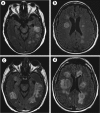
MRI images from Case 2. (a, b) Initial T2/fluid-attenuated inversion recovery (FLAIR) images demonstrated multiple intraparenchymal, supratentorial lesions situated predominantly in a periventricular distribution. These lesions were seen to exhibit punctate areas of susceptibility artifact, indicating a hemorrhagic component, as well as restricted diffusion (not pictured). (c, d) Five-day follow-up T2/FLAIR images demonstrated interval progression in the size and number of lesions, with new infratentorial lesions involving the cerebellum and cerebellar peduncles (not pictured). There was increased vasogenic edema and mass effect with evidence of entrapment of the temporal horn of the left lateral ventricle.

References

    1. Husain S, Muñoz P, Forrest G, Alexander BD, Somani J, Brennan K, Wagener MM, Singh N. Infections due to Scedosporium apiospermum and Scedosporium prolificans in transplant recipients: clinical characteristics and impact of antifungal agent therapy on outcome. Clin Infect Dis. 2005;40(1):89–99. - PubMed
    1. Tortorano AM, Richardson M, Roilides E, van Diepeningen A, Caira M, Munoz P, Johnson E, Meletiadis J, Pana ZD, Lackner M, Verweij P, Freiberger T, Cornely OA, Arikan-Akdagli S, Dannaoui E, Groll AH, Lagrou K, Chakrabarti A, Lanternier F, Pagano L, Skiada A, Akova M, Arendrup MC, Boekhout T, Chowdhary A, Cuenca-Estrella M, Guinea J, Guarro J, de Hoog S, Hope W, Kathuria S, Lortholary O, Meis JF, Ullmann AJ, Petrikkos G, Lass-Flörl C. ESCMID and ECMM joint guidelines on diagnosis and management of hyalohyphomycosis: Fusarium spp., Scedosporium spp. and others. Clin Microbiol Infect. 2014;20(Suppl 3):27–46. - PubMed
    1. Hartmann C, Müller C, Weißbrodt H, Suerbaum S, Tintelnot K, Stolle S, Hansen G, Sedlacek L. Successful prevention of scedosporiosis after lung transplantation in a cystic fibrosis patient by combined local and systemic triazole therapy. Med Mycol Case Rep. 2013;2:116–118. - PMC - PubMed
    1. Riddell J, 4th, Chenoweth CE, Kauffman CA. Disseminated Scedosporium apiospermum infection in a previously healthy woman with HELLP syndrome. Mycoses. 2004;47(9–10):442–446. - PubMed
    1. Marco de Lucas E, Sádaba P, Lastra García-Barón P, Ruiz Delgado ML, Cuevas J, Salesa R, Bermúdez A, González Mandly A, Gutiérrez A, Fernández F, Marco de Lucas F, Díez C. Cerebral scedosporiosis: an emerging fungal infection in severe neutropenic patients: CT features and CT pathologic correlation. Eur Radiol. 2006;16(2):496–502. - PubMed

Publication types

LinkOut - more resources