Review
. 2014 Jun 16:8:55.
doi: 10.3389/fncir.2014.00055. eCollection 2014.

Dopamine: a parallel pathway for the modulation of spinal locomotor networks

Affiliations
Review

Dopamine: a parallel pathway for the modulation of spinal locomotor networks

Simon A Sharples et al. Front Neural Circuits. .

Abstract

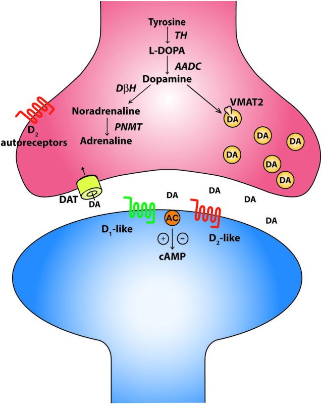
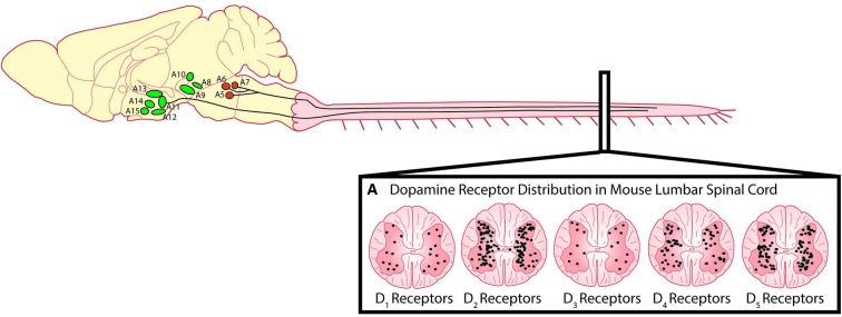
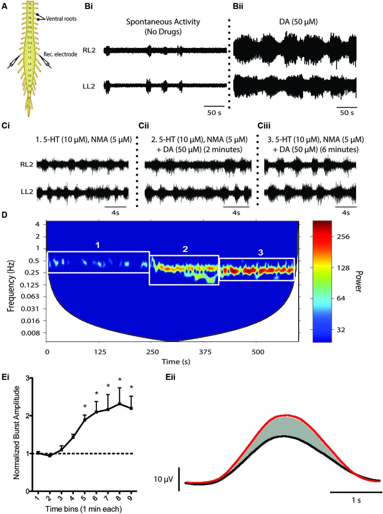
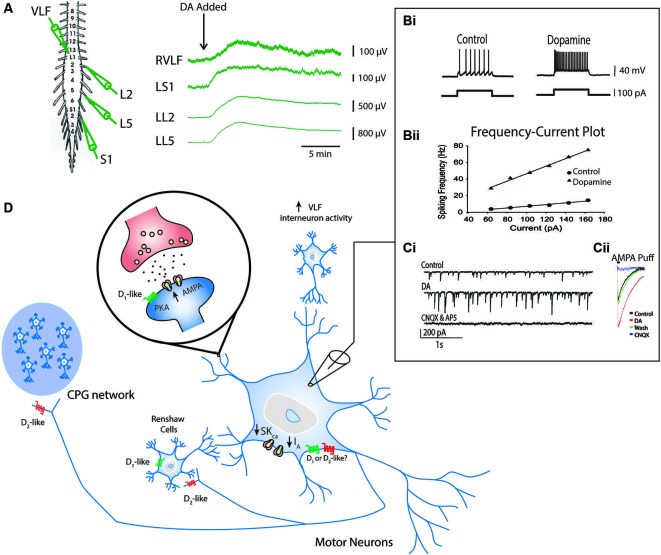
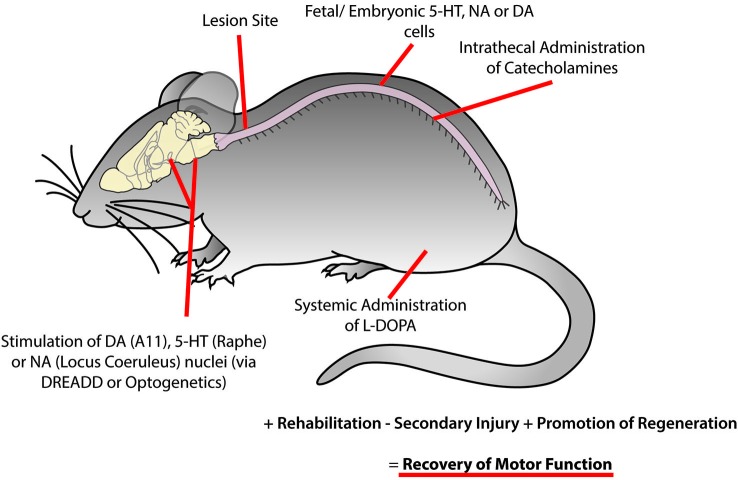
The spinal cord contains networks of neurons that can produce locomotor patterns. To readily respond to environmental conditions, these networks must be flexible yet at the same time robust. Neuromodulators play a key role in contributing to network flexibility in a variety of invertebrate and vertebrate networks. For example, neuromodulators contribute to altering intrinsic properties and synaptic weights that, in extreme cases, can lead to neurons switching between networks. Here we focus on the role of dopamine in the control of stepping networks in the spinal cord. We first review the role of dopamine in modulating rhythmic activity in the stomatogastric ganglion (STG) and the leech, since work from these preparations provides a foundation to understand its role in vertebrate systems. We then move to a discussion of dopamine's role in modulation of swimming in aquatic species such as the larval xenopus, lamprey and zebrafish. The control of terrestrial walking in vertebrates by dopamine is less studied and we review current evidence in mammals with a focus on rodent species. We discuss data suggesting that the source of dopamine within the spinal cord is mainly from the A11 area of the diencephalon, and then turn to a discussion of dopamine's role in modulating walking patterns from both in vivo and in vitro preparations. Similar to the descending serotonergic system, the dopaminergic system may serve as a potential target to promote recovery of locomotor function following spinal cord injury (SCI); evidence suggests that dopaminergic agonists can promote recovery of function following SCI. We discuss pharmacogenetic and optogenetic approaches that could be deployed in SCI and their potential tractability. Throughout the review we draw parallels with both noradrenergic and serotonergic modulatory effects on spinal cord networks. In all likelihood, a complementary monoaminergic enhancement strategy should be deployed following SCI.

Keywords: central pattern generator; dopamine; locomotion; monoamines; spinal cord.


Figures

Figure 1
Figure 1
The canonical catecholaminergic neuron is one that generates dopamine, noradrenaline or adrenaline. Catecholamines are synthesized from tyrosine through a series of biosynthetic steps progressing from tyrosine hydroxylase (TH) producing L-DOPA, aromatic amino acid decarboxylase (AADC) producing dopamine, dopamine-β-hydroxylase (DBH) producing noradrenaline and phenylethanolamine-N-methyl-transferase (PNMT) producing adrenaline. These neurons also express the vesicular monoaminergic transporter (VMAT2) to pump catecholamines into synaptic vesicles. A canonical dopaminergic neuron will also express dopamine reuptake transporters (DAT) and inhibitory D2 autoreceptors to regulate presynaptic release of dopamine. Post synaptic targets of dopamine include the excitatory D1-like and inhibitory D2-like receptors that act through G-protein second messenger cascades, increasing and decreasing intracellular cAMP levels respectively.
Figure 2
Figure 2
Descending dopaminergic fibres within the spinal cord originate in the A11. (A) Dopamine acts on all five dopamine receptors that are distributed non-uniformly through the dorsal and ventral horns of the spinal cord (Adapted with permission from Zhu et al., 2007). Descending noradrenergic fibres originating in the A5, A6 and A7 nuclei of the pons innervate the spinal cord. Schematic adapted with permission from Björklund and Dunnett (2007).
Figure 3
Figure 3
Dopaminergic modulation of spinal CPG activity. (A) Schematic of an in vitro isolated neonatal mouse spinal cord showing ventral root neurograms from the left and right L2 segment. (B) Spontaneous activity (Bi) is converted to a rhythmic slow non-locomotor rhythmic pattern (Bii) following bath application of dopamine (DA). (C and D) When DA is bath-applied during ongoing locomotor activity elicited by 5-HT and NMA it stabilizes and reduces the frequency of the rhythm. The spectrogram (D) depicts a cross-wavelet analysis of a locomotor rhythm evoked by 5-HT and NMA (box 1) and effect of dopamine (DA) on the pre-existing rhythm (box 2 and 3). Rhythm frequency is displayed on the y-axis and rhythm power displayed as warm or cool colors with warmer colors representing higher power or more stable rhythm. Dopamine also increases burst amplitude (Ei and Eii). (Ei) Graph displays an increase in burst amplitude over a 10 min period immediately following the addition of dopamine and (Eii) shows an average L2 neurogram burst from a representative experiment with bursts evoked by 5-HT and NMA (black) and following addition of dopamine (red) (Adapted from Humphreys and Whelan, 2012).
Figure 4
Figure 4
Known effects of dopamine on cellular components of the locomotor network. Ventral root neurograms from the L2, L5 and S1 segments and ventrolateral funiculus (VLF) of a spinal cord isolation preparation with fast synaptic transmission blocked (CNQX, AP5, PTX and Strychnine) indicates that dopamine increases the excitability of motor neurons projecting through ventral roots and interneurons projecting through the VLF (A) (Adapted from Han et al., 2007). Intracellular recordings from motor neurons show that dopamine increases motor neuron excitability indicated by an increase in the slope of the frequency-current relationship (Bi and Bii). This effect is in part mediated by a reduction in IA and SKCa conductances. Dopamine also increases AMPA conductances (Ci and Cii) via a D1-like receptor mechanisms (Adapted with permission from Han and Whelan, 2009). Dopamine reduces recurrent excitatory feedback to the locomotor CPG (Humphreys and Whelan, 2012) and Renshaw cells via D2-like receptor mechanisms (Maitra et al., 1993). A summary of all these effects are depicted in panel (D).
Figure 5
Figure 5
Recovery of motor function following spinal cord injury may be facilitated using combinatorial therapies to promote plasticity within motor networks. This could be accomplished by targeting monoaminergic systems through the activation of descending brain nuclei using optogenetic or pharmacogenetic approaches, implantation of fetal or embryonic cells from the dopaminergic ventral tegmental area, noradrenergic locus coeruleus or serotonergic raphe nucleus caudal to the site of injury, intrathecal injection of catecholamines and systemic injection of L-DOPA. While these approaches may not be optimal in isolation, they may serve as an effective complementary treatment with rehabilitation and other therapies to reduce secondary injury and promote regeneration of damaged descending tracts to promote the recovery of motor function.

References

    1. Abrahamson E. E., Moore R. Y. (2001). The posterior hypothalamic area: chemoarchitecture and afferent connections. Brain Res. 889, 1–22 10.1016/s0006-8993(00)03015-8 - DOI - PubMed
    1. Airan R. D., Thompson K. R., Fenno L. E., Bernstein H., Deisseroth K. (2009). Temporally precise in vivo control of intracellular signalling. Nature 458, 1025–1029 10.1038/nature07926 - DOI - PubMed
    1. Aston-Jones G., Deisseroth K. (2013). Recent advances in optogenetics and pharmacogenetics. Brain Res. 1511, 1–5 10.1016/j.brainres.2013.01.026 - DOI - PMC - PubMed
    1. Baker L. L., Chandler S. H., Goldberg L. J. (1984). L-dopa-induced locomotor-like activity in ankle flexor and extensor nerves of chronic and acute spinal cats. Exp. Neurol. 86, 515–526 10.1016/0014-4886(84)90086-4 - DOI - PubMed
    1. Barbeau H., Rossignol S. (1987). Recovery of locomotion after chronic spinalization in the adult cat. Brain Res. 412, 84–95 10.1016/0006-8993(87)91442-9 - DOI - PubMed

Publication types