. 2014 Jun 17:7:66.
doi: 10.1186/1757-2215-7-66. eCollection 2014.

Expression of the erythropoietin receptor by germline-derived cells - further support for a potential developmental link between the germline and hematopoiesis

Affiliations

Expression of the erythropoietin receptor by germline-derived cells - further support for a potential developmental link between the germline and hematopoiesis

Malwina Suszynska et al. J Ovarian Res. .

Abstract

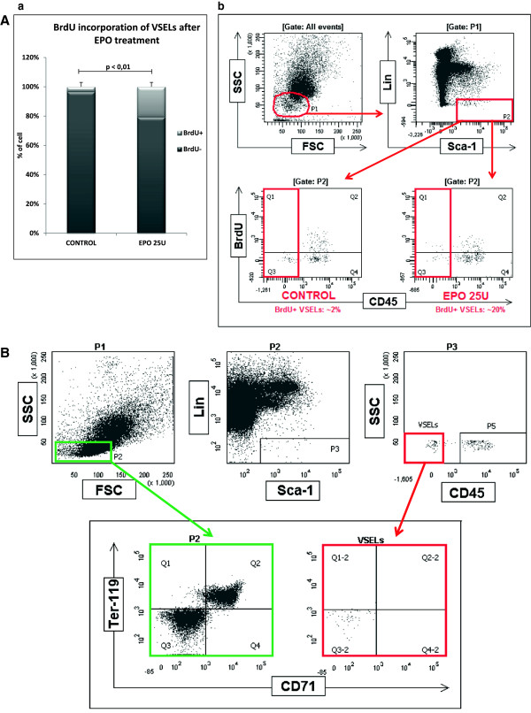
Background: Expressing several markers of migrating primordial germ cells (PGCs), the rare population of quiescent, bone marrow (BM)-residing very small embryonic-like stem cells (VSELs) can be specified like PGCs into hematopoietic stem/progenitor cells (HSPCs). These two properties of VSELs support the possibility of a developmental origin of HSPCs from migrating PGCs.

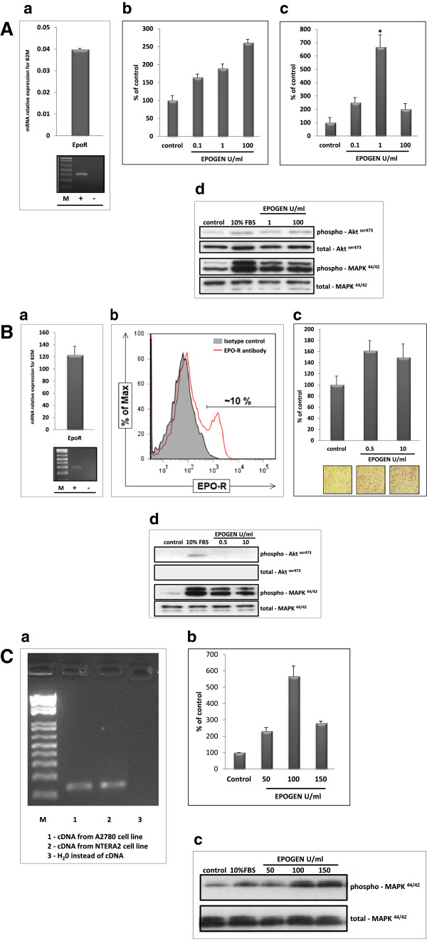
Methods: To address a potential link between VSELs and germ line cells we analyzed by RT-PCR and FACS expression of erythropoietin receptor (EpoR) on murine bone marrow- and human umbilical cord blood-derived VSELs, murine and human teratocarcinoma cell lines and human ovarian cancer cells. A proper gating strategy and immunostaining excluded from FACS analysis potential contamination by erythroblasts. Furthermore, the transwell chemotaxis assays as well as adhesion and signaling studies were performed to demonstrate functionality of erythropoietin - EpoR axes on these cells.

Results: We report here that murine and human VSELs as well as murine and human teratocarcinoma cell lines and ovarian cancer cell lines share a functional EpoR.

Conclusions: Our data provide more evidence of a potential developmental link between germline cells, VSELs, and HSCs and sheds more light on the developmental hierarchy of the stem cell compartment in adult tissues.

Keywords: Cancer development; EpoR; Germ line; Ovarian cancer; VSELs.


Figures

Figure 1
Figure 1
Erythropoietin induces proliferation of quiescent BM VSELs. Panel A. BrdU incorporation into VSELs after EPO treatment. (a) The percentage of VSELs that incorporated BrdU into newly synthesized DNA. After treatment with 10 doses of EPO at 25 U/dose, ~20% of VSELs are BrdU+, in contrast to the control group (~2% of VSELs are BrdU+). (b) Incorporation of BrdU was measured by FACS. Representative FACS analysis of six experiments is shown. Panel B. Analysis of VSELs for the presence of erythroid markers. Cells were fixed and stained with 7AAD to show only events exhibiting DNA content, gate P1 (not shown). Gate P2 includes small, agranular cells. Gate P3 includes Sca-1+/Lin- cells, which are visualized on the next dot plot as CD45-negative cells (VSELs) and CD45-positive cells (HSPCs). Erythroblast markers CD 71 and Ter-119 are not expressed on VSELs (right, lower panel) in contrast to control cells, where some of the cells express CD71 and Ter-119 among the population from the P2 gate (left, lower panel). One representative dot plot analysis out of three is shown.
Figure 2
Figure 2
EpoR is expressed by murine and human VSELs. Panel A. RQ-PCR results for EpoR expression by purified murine (left panel) and human (right panel) VSELs, HSCs, and MNCs. Combined data from three independent experiments are shown. Panel B. Immunostaining of EpoR and Oct4 protein in purified murine VSELs (left panel) and HSCs (right panel). The images were acquired using an Olympus FV1000 confocal microscope. A representative staining is shown. The experiment was performed on three independent cell sorts. Panel C. Detection of EpoR expression by FACS among hCB-derived VSELs. The left panel presents the percentages of CD34+ and CD133+ VSELs that express EpoR. The level of expression of EpoR on UCB-derived VSELs is ~20% for the population of Lin-/CD45-/CD34+ VSELs and ~15% for the population of Lin-/CD45-/CD133+ VSELs. The right panel shows one representative set of results out of ten FACS analyses.
Figure 3
Figure 3
Functional EpoR is expressed on murine and human germline-derived immortalized cell lines. Panel A. (a) RT-PCR analysis of EpoR expression in the P19 murine teratocarcinoma cell line. Upper panel, RQ-PCR data, lower panel, SDS-PAGE gel of RT-PCR amplicon. The experiment was performed twice with similar results. (b). The effect of erythropoietin (0–100 U/ml) on adhesion of P19 cells to fibronectin. Combined data from three independent experiments are shown. (c). The effect of erythropoietin (0–100 U/ml) on the chemoattraction of P19 cells across Transwell membranes covered with gelatin. Combined data from three independent experiments are shown. (d). Phosphorylation of AKT and p42/44 MAPK in P19 cells. A representative blot is shown. Panel B. (a) RT-PCR analysis of EpoR expression in the NTERA2 human teratocarcinoma cell line. Upper panel, RQ-PCR data; lower panel, SDS-PAGE gel of RT-PCR amplicon. Experiment was performed twice with similar results. (b). Expression of EpoR on the NTERA2 cell line as measured by FACS. One representative histogram out of three experiments is shown. (c). Effect of erythropoietin (0–10 U/ml) on adhesion of NTERA2 cells to fibronectin. Combined data from three independent experiments are shown. (d). Phosphorylation of AKT and p42/44 MAPK in NTERA2 cells. One representative blot out of two is shown. Panel C. (a) RT-PCR analysis of EpoR expression in the A2780 human ovarian cancer cell line (lane 1). Lane 2, positive control (NTERA2 teratocarcinoma cell line). Lane 3, negative control (H2O instead of cDNA). The experiment was repeated twice with similar results. (b). Effect of erythropoietin on chemoattraction of the A2780 ovarian cancer cell line across Transwell membranes. Combined data from two independent experiments are shown. (c). Phosphorylation of p42/44 MAPK in the A2780 human ovarian cancer cell line stimulated by erythropoietin. A representative blot is shown.

Similar articles

Cited by

References

    1. Suszynska M, Zuba-Surma EK, Maj M, Mierzejewska K, Ratajczak J, Kucia M, Ratajczak MZ. The proper criteria for identification and sorting of very small embryonic-like stem cells, and some nomenclature issues. Stem Cells Dev. 2014;23:702–713. - PMC - PubMed
    1. Ohtaka T, Matsui Y, Obinata M. Hematopoietic development of primordial germ cell-derived mouse embryonic germ cells in culture. Biochem Biophys Res Com. 1999;260:475–482. - PubMed
    1. Rich IN. Primordial germ cells are capable of producing cells of the hematopoietic system in vitro. Blood. 1995;86:463–472. - PubMed
    1. Yoshimoto M, Heike T, Chang H, Kanatsu-Shinohara M, Baba S, Varnau JT, Shinohara T, Yoder MC, Nakahata T. Bone marrow engraftment but limited expansion of hematopoietic cells from multipotent germline stem cells derived from neonatal mouse testis. Exp Hematol. 2009;37:1400–1410. - PMC - PubMed
    1. Kritzenberger M, Wrobel KH. Histochemical in situ identification of bovine embryonic blood cells reveals differences to the adult haematopoietic system and suggests a close relationship between haematopoietic stem cells and primordial germ cells. Histochem Cell Biol. 2004;121:273–289. - PubMed

Publication types