Review
. 2014 Jun 7;4(4):365-84.
eCollection 2014.

PET radiopharmaceuticals for imaging of tumor hypoxia: a review of the evidence

Affiliations
Review

PET radiopharmaceuticals for imaging of tumor hypoxia: a review of the evidence

Egesta Lopci et al. Am J Nucl Med Mol Imaging. .

Abstract

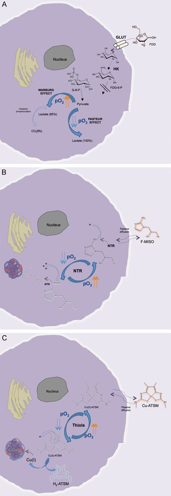
Hypoxia is a pathological condition arising in living tissues when oxygen supply does not adequately cover the cellular metabolic demand. Detection of this phenomenon in tumors is of the utmost clinical relevance because tumor aggressiveness, metastatic spread, failure to achieve tumor control, increased rate of recurrence, and ultimate poor outcome are all associated with hypoxia. Consequently, in recent decades there has been increasing interest in developing methods for measurement of oxygen levels in tumors. Among the image-based modalities for hypoxia assessment, positron emission tomography (PET) is one of the most extensively investigated based on the various advantages it offers, i.e., broad range of radiopharmaceuticals, good intrinsic resolution, three-dimensional tumor representation, possibility of semiquantification/quantification of the amount of hypoxic tumor burden, overall patient friendliness, and ease of repetition. Compared with the other non-invasive techniques, the biggest advantage of PET imaging is that it offers the highest specificity for detection of hypoxic tissue. Starting with the 2-nitroimidazole family of compounds in the early 1980s, a great number of PET tracers have been developed for the identification of hypoxia in living tissue and solid tumors. This paper provides an overview of the principal PET tracers applied in cancer imaging of hypoxia and discusses in detail their advantages and pitfalls.

Keywords: 18F-FAZA; 18F-FDG; 18F-FMISO; 64Cu-ATSM; Hypoxia; PET; tumor imaging.


Figures

Figure 1
Figure 1
Overview of the uptake and retention mechanisms of FDG (A), F-MISO (B), and Cu-ATSM (C) in living cells under hypoxic conditions. For FDG there is a wide overlap between the cellular uptake in normoxic (Warburg effect) and hypoxic conditions (Pasteur effect). For the other two tracers, after passive diffusion through the membrane, the radiopharmaceutical is retained according to the oxygen tension (pO2) present in the intracellular environment: in the presence of reduced pO2, F-MISO undergoes progressive reduction by the nitroreductase enzyme (NTR); also, Cu(II)-ATSM nuclide is reduced to copper (I) by the intracellular thiols, making the Cu-ATSM complex less stable. Both processes are reversible in the presence of sufficient O2, and the molecules (F-MISO and Cu (II)-ATSM) are free to leave the cell. Conversely, in hypoxic conditions the Cu(I)-ATSM complex is progressively dissociated, with the formation of H2-ATSM and free Cu(I), which is very rapidly incorporated into intracellular proteins. In contrast, the reduced F-MISO is covalently bound to the intracellular proteins [59,84,122,128]. GLUT, glucose transporter; HK, hexokinase; G-6-P, glucose-6-phosphate).
Figure 2
Figure 2
Example of a patient with localized head and neck squamous cell carcinoma (HNSCC) who was investigated with 18F-FDG PET before (A) and after the end of RT (B). At staging the patient had undergone 64Cu-ATSM PET/CT (C-E) documenting some mild uptake at the level of the primary tumor in the left tonsil (SUVmax 1.85). As is visible in (B) the patient achieved a complete response after treatment.
Figure 3
Figure 3
Example of a patient with advanced HNSCC who was investigated with 18F-FDG PET at staging (A) and after the end of combined chemoradiotherapy (B). Before treatment the patient underwent 64Cu-ATSM PET/CT (C-E), documenting intense tracer uptake (SUVmax 17.86) both in the primary tumor, involving the right tonsil, and in numerous bilateral cervical nodes. Despite the high-dose therapeutic regimen utilized, the patient presented some residual disease at end-of-treatment evaluation (B), as confirmed during follow-up.

References

    1. Vaupel P, Mayer A. Hypoxia in cancer: significance and impact on clinical outcome. Cancer Metastasis Rev. 2007;26:225–239. - PubMed
    1. Tomlinson RH, Gray LH. The histological structure of some human lung cancers and the possible implications for radiotherapy. Br J Cancer. 1955;9:539–49. - PMC - PubMed
    1. Mees G, Dierckx R, Vangestel C, Van de Wiele C. Molecular imaging of hypoxia with radiolabelled agents. Eur J Nucl Med Mol Imaging. 2009;36:1674–1686. - PMC - PubMed
    1. Anderson CJ, Ferdani R. Copper-64 radiopharmaceuticals for PET imaging of cancer: advances in preclinical and clinical research. Cancer Biother Radiopharm. 2009;24:379–393. - PMC - PubMed
    1. Albiach CF, Moreno AC, Cordon MR, Morillo Macías V, Bouché Babiloni A, Beato Tortajada I, Sánchez Iglesias A, Francés Muñoz A. Contribution of hypoxia-measuring molecular imaging techniques to radiotherapy planning and treatment. Clin Transl Oncol. 2010;12:22–26. - PubMed

LinkOut - more resources