Review
. 2014 Sep 17;5(9):776-83.
doi: 10.1021/cn5000666. Epub 2014 Jul 10.

A novel restricted diffusion model of evoked dopamine

Affiliations
Review

A novel restricted diffusion model of evoked dopamine

Seth H Walters et al. ACS Chem Neurosci. .

Abstract

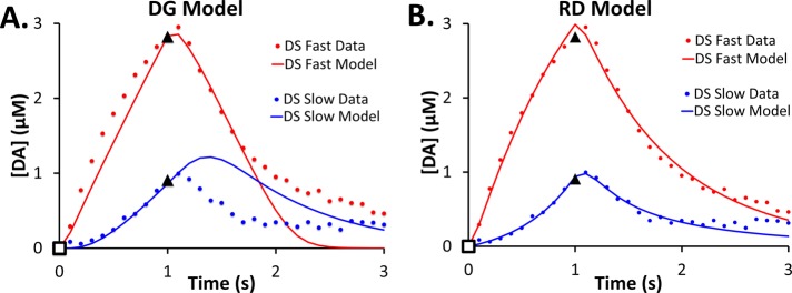
In vivo fast-scan cyclic voltammetry provides high-fidelity recordings of electrically evoked dopamine release in the rat striatum. The evoked responses are suitable targets for numerical modeling because the frequency and duration of the stimulus are exactly known. Responses recorded in the dorsal and ventral striatum of the rat do not bear out the predictions of a numerical model that assumes the presence of a diffusion gap interposed between the recording electrode and nearby dopamine terminals. Recent findings, however, suggest that dopamine may be subject to restricted diffusion processes in brain extracellular space. A numerical model cast to account for restricted diffusion produces excellent agreement between simulated and observed responses recorded under a broad range of anatomical, stimulus, and pharmacological conditions. The numerical model requires four, and in some cases only three, adjustable parameters and produces meaningful kinetic parameter values.

Keywords: Model; diffusion; domain; dopamine; restricted diffusion; voltammetry.


Figures

Figure 1
Figure 1
(A) Evoked responses, as predicted by eq 1 (red line), rise during the stimulus and decay back to zero after the stimulus ends. However, observed responses (green line) also exhibit lag (an initial delay in the appearance of the signal), overshoot (the signal continues to rise after the stimulus ends), and hang-up (the signal remains elevated for prolonged periods after the stimulus ends instead of returning to baseline). The open square indicates the start of the stimulus, and the closed triangles indicate the end of the stimulus. (B) Schematic representation of the RD model (see the Methods section for definitions of the parameters). The extracellular space is divided into inner (IC) and outer (OC) compartments. DA is released from axon terminals (at) to the IC, is subsequently transported to the OC, and is removed from the OC by uptake. The model postulates that FSCV recording takes place in the OC.
Figure 2
Figure 2
(A) Evoked responses recorded in fast domains of the DS and NAc (stimulus = 200 ms, 60 Hz, 250 μA): the solid lines are the averaged responses, and the dotted lines are the SEM intervals. (B) DG simulations using region-specific parameter values and Gap values of 1 and 5. (C) DG simulations of the averaged DS and NAc data points (SEMs omitted for clarity). (D) RD simulations of the averaged DS and NAc data points (SEMs omitted for clarity). The open square indicates when the stimulus begins, and the closed triangle marks the data point at the end of the stimulus. The parameter values are reported in the Supporting Information.
Figure 3
Figure 3
Fits of the DG (A) and RD (B) models to averaged responses from fast and slow domains of the dorsal striatum. The parameter values for these fits are reported in the Supporting Information.
Figure 4
Figure 4
Fits of the DG (A) and RD (B) models to averaged responses from the dorsal striatum (A, B) and nucleus accumbens (B). In panel A, “predrug” refers to the stimulus as collected at a recording site in a drug naive rat, whereas “nomifensine” refers to data collected at the same site after i.p. administration of the competitive uptake inhibitor nomifensine. The parameter values are reported in the Supporting Information.
Figure 5
Figure 5
Fits of the RD model to averaged responses from the nucleus accumbens (A, B) and dorsal striatum (C, D) both before (blue) and after (green) animals were treated with nomifensine. The parameter values are listed in the Supporting Information.
Figure 6
Figure 6
Three-parameter RD simulations of postnomifensine averaged responses to 0.2 s, 60 Hz stimuli recorded in the dorsal striatum and the nucleus accumbens. Parameter values are reported in Table 2.

References

    1. Schultz W. (2007) Multiple dopamine functions at different time courses. Annu. Rev. Neurosci. 30, 259–288. - PubMed
    1. Abi-Dargham A.; Rodenhiser J.; Printz D.; Zea-Ponce Y.; Gil R.; Kegeles L. S.; et al. (2000) Increased baseline occupancy of D2 receptors by dopamine in schizophrenia. Proc. Natl. Acad. Sci. U.S.A. 97, 8104–8109. - PMC - PubMed
    1. Schultz W. (1982) Depletion of dopamine in the striatum as an experimental model of Parkinsonism: direct effects and adaptive mechanisms. Prog. Neurobiol. (Oxford, U.K.) 18, 121–166. - PubMed
    1. Sagvolden T.; Johansen E. B.; Aase H.; Russell V. A. (2005) A dynamic developmental theory of attention-deficit/hyperactivity disorder (ADHD) predominantly hyperactive/impulsive and combined subtypes. Behav. Brain Sci. 28, 397–419. - PubMed
    1. Carlsson A.; Lindqvist M.; Magnusson T.; Waldeck B. (1958) On the presence of 3-hydroxytyramine in brain. Science 127, 471. - PubMed

Publication types

LinkOut - more resources