. 2014 Jul 1;9(7):e100639.
doi: 10.1371/journal.pone.0100639. eCollection 2014.

An unstable Th epitope of P. falciparum fosters central memory T cells and anti-CS antibody responses

Affiliations

An unstable Th epitope of P. falciparum fosters central memory T cells and anti-CS antibody responses

Carlos A Parra-López et al. PLoS One. .

Erratum in

  • PLoS One. 2014;9(11):e113028. Yin, Liusong [added]

Abstract

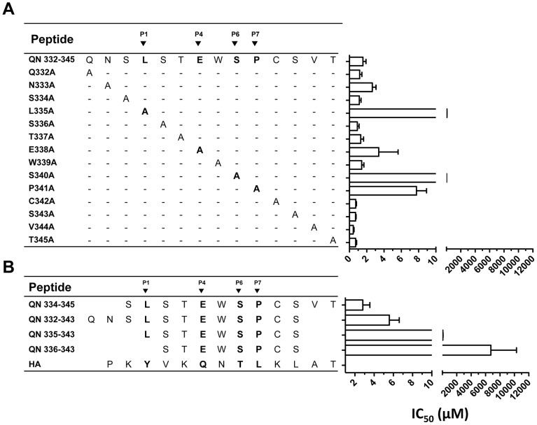
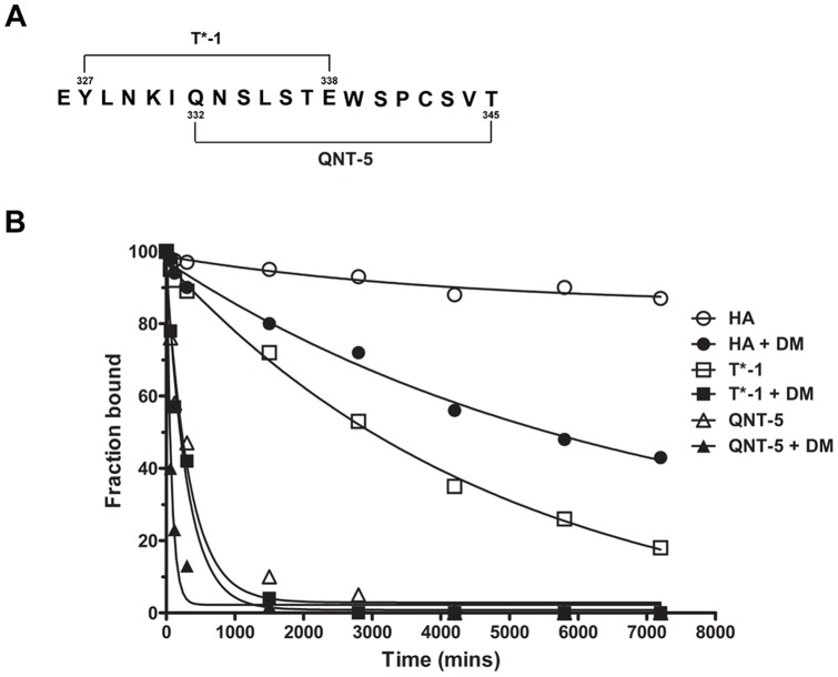
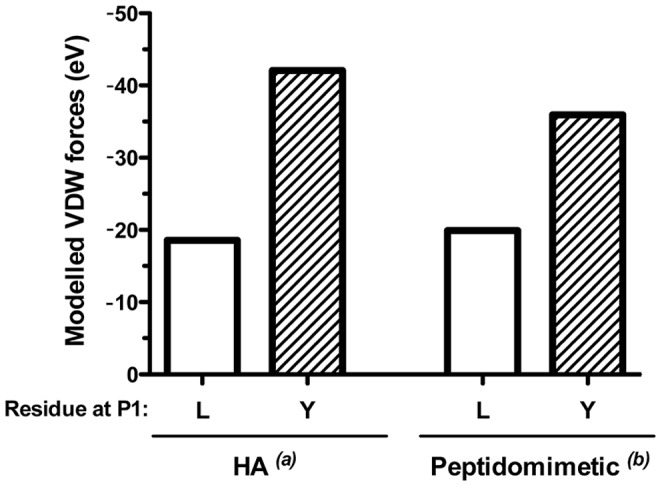
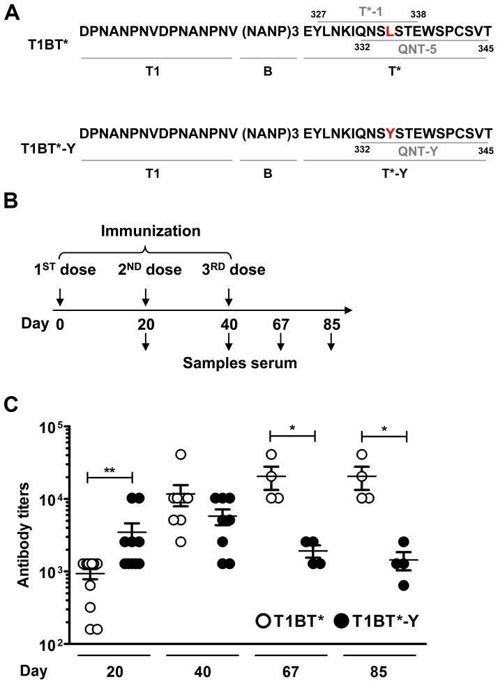
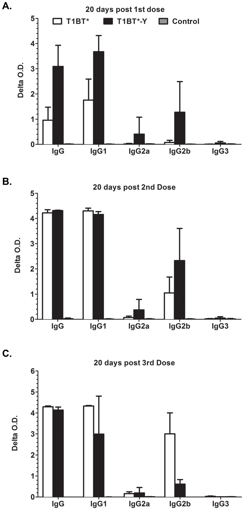
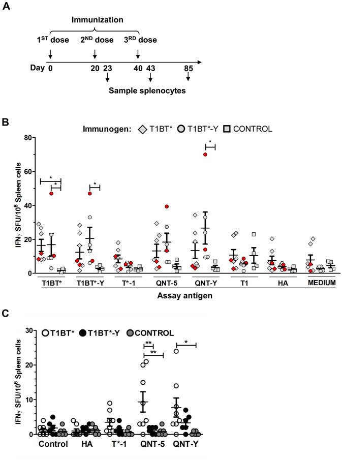
Malaria is transmitted by Plasmodium-infected anopheles mosquitoes. Widespread resistance of mosquitoes to insecticides and resistance of parasites to drugs highlight the urgent need for malaria vaccines. The most advanced malaria vaccines target sporozoites, the infective form of the parasite. A major target of the antibody response to sporozoites are the repeat epitopes of the circumsporozoite (CS) protein, which span almost one half of the protein. Antibodies to these repeats can neutralize sporozoite infectivity. Generation of protective antibody responses to the CS protein (anti-CS Ab) requires help by CD4 T cells. A CD4 T cell epitope from the CS protein designated T* was previously identified by screening T cells from volunteers immunized with irradiated P. falciparum sporozoites. The T* sequence spans twenty amino acids that contains multiple T cell epitopes restricted by various HLA alleles. Subunit malaria vaccines including T* are highly immunogenic in rodents, non-human primates and humans. In this study we characterized a highly conserved HLA-DRβ1*04:01 (DR4) restricted T cell epitope (QNT-5) located at the C-terminus of T*. We found that a peptide containing QNT-5 was able to elicit long-term anti-CS Ab responses and prime CD4 T cells in HLA-DR4 transgenic mice despite forming relatively unstable MHC-peptide complexes highly susceptible to HLA-DM editing. We attempted to improve the immunogenicity of QNT-5 by replacing the P1 anchor position with an optimal tyrosine residue. The modified peptide QNT-Y formed stable MHC-peptide complexes highly resistant to HLA-DM editing. Contrary to expectations, a linear peptide containing QNT-Y elicited almost 10-fold lower long-term antibody and IFN-γ responses compared to the linear peptide containing the wild type QNT-5 sequence. Some possibilities regarding why QNT-5 is more effective than QNT-Y in inducing long-term T cell and anti-CS Ab when used as vaccine are discussed.


Conflict of interest statement

Competing Interests: The authors have declared that no competing interests exist.

Figures

Figure 1
Figure 1. Identification of residues in QNT-5 critical for binding to DR4.
A series of QNT-5 analogues with single alanine substitutions (A) and N- and C-terminal truncated QNT-5 peptides (B) were synthesized and tested at concentrations ranging from 0 to 100 µM for their ability to bind to DR4. Relative binding was measured in a binding inhibition assay. The concentration of each alanine analogue or truncated peptide required to reduce the binding of a biotin-labeled test peptide to 50% (IC50) is shown. The test peptide was a biotinylated version of HA (306PKYVKQNTLKLAT318), a peptide from influenza hemagglutinin that binds strongly to DR4. Under the conditions used in this set of experiments, the IC50 for the HA peptide was 0.31±0.05 µM (Table S1). Representative findings from 3 experiments performed are shown (each point was performed in duplicate). Bars represent SD.
Figure 2
Figure 2. Dissociation kinetics from DR4 of HA, T*-1 and QNT-5 peptides measured in presence and absence of HLA-DM.
(A) Amino-acid sequence and location of T*-1 and QNT-5 epitopes in T*. (B) Characterization of the dissociation behavior of peptide MHC complexes formed after 72 h of complex formation. The curves shown represent single or double exponential decays that fit the data. Filled symbols represent the decays values of the 3 DR4-peptide complexes in the presence of HLA-DM. Empty symbols represents the decay values of the complexes in the absence of HLA-DM. A representative experiment from 2 experiments performed is shown (each time point was carried out in duplicate).
Figure 3
Figure 3. VDW energy value for the hydrophobic interactions at P1 between DR4 and QNT-5 or an analogue of QNT-5 with a L335Y substitution at P1 (peptide QNT-Y).
VDW value after the computational docking of peptides QNT-5 and QNT-Y into an MHC (DRβ1*04:01:01) structure, originally determined for the HA peptide and peptidomimetic inhibitor complexes, with a tyrosine(a) (left panel) and a cyclohexylalanine(b) side chain at P1 (right panel), respectively. The height of the white- and dashed-boxes represents the VDW free energy value expressed in eV for QNT-5 (L) with the anchor residue L335 at P1 and for QNT-Y (Y) with Y318 at P1, respectively.
Figure 4
Figure 4. Binding activity of QNT-Y and stability of the DR4/QNT-Y peptide complex.
(A) Amino-acid sequence of QNT-5 and QNT-Y peptide analogue. (B) Competition binding assay for QNT-5, QNT-Y and HA. The plots show the binding inhibition of the biotinylated HA306–318 peptide to DR4 using increasing amounts (0 to 20 µM) of unlabeled peptides to calculate the concentration of each peptide required to reduce the binding of a biotin-labeled test peptide to 50% (IC50). Under the experimental conditions used here, the IC50 for the HA peptide was ∼172 µM. Representative results from 1 of 3 experiments performed are shown (each point was performed in duplicate). (C) Dissociation kinetics of the QNT-5 and QNT-Y peptides from DR4, measured in the presence (filled symbols) or absence (empty) of HLA-DM. A representative experiment of 3 performed is shown (each time point was performed in duplicate). Bars represent SEM.
Figure 5
Figure 5. Long term quantitation of anti-(NANP)6 Ab responses by ELISA in mice vaccinated with T1BT* or T1BT*-Y peptides.
(A) Amino-acid sequences of T1BT* and T1BT*-Y polypeptides used for vaccination of HLA-DR4 transgenic mice. T1BT* comprise T1 (a T cell epitope from the 5′minor repeat region of P. falciparum CS protein [31]), B (three copies of immune-dominant B-cell repeat epitope (NANP) from P. falciparum CS protein [97]) and the NF54 variant of T* epitope that includes T*-1327–338 YLNKIQNSLSTE and QNT-5332–345 QNSLSTEWSPCSVT with L335 highlighted in red. T1BT*-Y is identical to T1BT* except that QNT-5332–345 QNSYSTEWSPCSVT harbors the single amino-acid substitution L335Y (in red). (B) Immunization scheme indicating the days when serum samples were collected. (C) Anti-(NANP)6 antibody titers in serum samples of mice immunized with T1BT* (open) or T1BT*-Y (black circles) during the course of the immunization protocol. The mean anti-(NANP)6 Ab titers correspond to the average titer determined in the sera of groups of 3 DR4 transgenic mice immunized with T1BT* or T1BT*-Y in 3 independent experiments. (*) p<0.05; (**) p<0.001 Mann Whitney test, mean with SEM (standard errors of the mean) bars are shown.
Figure 6
Figure 6. IgG Isotype responses in T1BT* and T1BT*-Y immunized mice.
IgG subtype of anti-(NANP)6 antibody responses elicited in DR4 transgenic mice twenty days after the first (A), second (B) and third dose (C) of T1BT* (white bars); T1BT*-Y (black bars) peptides or Montanide ISA 720 (grey bars). The bars indicate mean delta O.D. (optical density serum in wells coated with (NANP)6 minus PBS wells) obtained with DR4 transgenic serum (1∶80 dilution) incubated with (NANP)6 peptide-coated ELISA plates and reacted with IgG subtype-specific antibodies. Serum samples were tested individually and means and standard deviation for the group are shown.
Figure 7
Figure 7. Quantitation of IFN-γ secreting cells in the spleens of mice after vaccination with T1BT* or T1BT*-Y by ELISPOT.
(A) Immunization scheme indicating the days when splenocytes for ELISPOT were collected. (B) The graph shows the mean number of splenocytes producing IFN-γ per 1×10⁶ cells from mice immunized with T1BT* (diamonds), T1BT*-Y (filled circles) or adjuvant/PBS (squares) after stimulation for 48 h in vitro with the assay antigens (T1BT*, T1BT*-Y, T*-1, QNT-5, QNT-Y, T1 and HA (10 µg/mL each)). The p values are relative to control mice immunized with PBS/adjuvant; * p<0.05. Kruskal-Wallis test with Dunn's Multiple Comparison Test. The IFN-γ SFU at day 20 from mice immunized with only 2 antigen doses is shown in red. Mean with SEM (standard error of the mean) bars are shown. (C) IFN-γ secreting cells quantified by ELISPOT at day 85 in spleens of DR4 transgenic mice immunized with T1BT* (open circles), T1BT*-Y (black) or adjuvant/PBS (gray) after stimulation for 48 h in vitro with media (control) or the assay antigens HA; T*-1, QNT-5, QNT-Y (10 µg/mL each). (*) p<0.05; (**) p<0.001 Kruskal-Wallis test with Dunn's Multiple Comparison Test. Mean with SEM bars are shown.
Figure 8
Figure 8. Human CD4 T cells specific for the QNT-5 epitope cross-react with QNT-Y and vice versa.
(A) Prime-boost scheme used for the in vitro priming of naïve CD4 T cells with T1BT* or T1BT*-Y pulsed DCs from a DRβ1*04:01:01 healthy individual. (B) Flow cytometric analysis of CD4 naïve T cells following co-culture for 14 days with DCs pulsed with T1BT* (top) or T1BT*-Y (bottom) and re-stimulated with peptide-pulsed DCs for 6 days. Plots show the percentages of CD4 T cells stained with SA-PE QNT-5, with SA-PE QNT-Y tetramers or with SA-PE only among CD3+CD4+ T cells after 21 days in culture. (C) CD4+ naïve (TN) (CD45RO- CD62-L+), central memory (TCM) (CD45RO+ CD62-L+), effector (TEM) (CD45RO+ CD62-L-) and terminal effector (TEMRA) (CD45RO- CD62-L-) T cell sub-populations were identified using flow cytometry by staining the cells after 21 days in culture with fluorescently labeled anti-CD45RO and anti-CD62-L antibodies. The numbers in the plots correspond to the percentage of each sub-population among CD3+CD4+ double-positive T cells. (D) Plots showing the percentages of CD4 T cells positive for QNT-5 and QNT-Y tetramers present among the TN, TCM, TEM or TEMRA T cell sub-populations after 21 days in culture.

Similar articles

Cited by

References

    1. WHO (2011) WORLD HEALTH STATISTICS 2011. WHO Library Cataloguing-in-Publication Data.
    1. Nussenzweig RS, Vanderberg J, Most H, Orton C (1967) Protective immunity produced by the injection of x-irradiated sporozoites of plasmodium berghei. Nature 216: 160–162. - PubMed
    1. Clyde DF (1975) Immunization of man against falciparum and vivax malaria by use of attenuated sporozoites. The American journal of tropical medicine and hygiene 24: 397–401. - PubMed
    1. Rieckmann KH (1990) Human immunization with attenuated sporozoites. Bulletin of the World Health Organization 68 Suppl13–16. - PMC - PubMed
    1. Hoffman SL, Goh LM, Luke TC, Schneider I, Le TP, et al. (2002) Protection of humans against malaria by immunization with radiation-attenuated Plasmodium falciparum sporozoites. The Journal of infectious diseases 185: 1155–1164. - PubMed

Publication types