. 2014 Jun 30;15(7):11637-64.
doi: 10.3390/ijms150711637.

Supraspliceosomes at defined functional states portray the pre-assembled nature of the pre-mRNA processing machine in the cell nucleus

Affiliations

Supraspliceosomes at defined functional states portray the pre-assembled nature of the pre-mRNA processing machine in the cell nucleus

Hani Kotzer-Nevo et al. Int J Mol Sci. .

Abstract

When isolated from mammalian cell nuclei, all nuclear pre-mRNAs are packaged in multi-subunit large ribonucleoprotein complexes-supraspliceosomes-composed of four native spliceosomes interconnected by the pre-mRNA. Supraspliceosomes contain all five spliceosomal U snRNPs, together with other splicing factors, and are functional in splicing. Supraspliceosomes studied thus far represent the steady-state population of nuclear pre-mRNAs that were isolated at different stages of the splicing reaction. To analyze specific splicing complexes, here, we affinity purified Pseudomonas aeruginosa phage 7 (PP7)-tagged splicing complexes assembled in vivo on Adenovirus Major Late (AdML) transcripts at specific functional stages, and characterized them using molecular techniques including mass spectrometry. First, we show that these affinity purified splicing complexes assembled on PP7-tagged AdML mRNA or on PP7-tagged AdML pre-mRNA are assembled in supraspliceosomes. Second, similar to the general population of supraspliceosomes, these defined supraspliceosomes populations are assembled with all five U snRNPs at all splicing stages. This study shows that dynamic changes in base-pairing interactions of U snRNA:U snRNA and U snRNA:pre-mRNA that occur in vivo during the splicing reaction do not require changes in U snRNP composition of the supraspliceosome. Furthermore, there is no need to reassemble a native spliceosome for the splicing of each intron, and rearrangements of the interactions will suffice.


Figures

Figure 1
Figure 1
Scheme of the affinity purification of specific supraspliceosomes. (A,B) Schemes of AdML constructs used for preparation of stable cell lines, each expressing one of the constructs used for the affinity purification of specific supraspliceosomes at defined splicing stages. (A) Constructs expressing AdML-WT; (B) constructs expressing AdML-Mut, having a mutated 3' splice site, designated “x”. The upper schemes represent the control construct without PP7 tag. The middle scheme of (B) and the lower scheme of (A) represent the constructs harboring the PP7 tag at the 3'UTR. The lower scheme of (B) represents the construct harboring the PP7 tag at the intron. Open boxes represent exons, lines represent introns, and stem-loops represent the PP7 tag; (C) Scheme of the affinity purification process of specific splicing complexes assembled on a specific transcript. First, nuclear supernatants enriched in splicing complexes (scheme in yellow) are extracted from HeLa cell-lines expressing the respective AdML construct. A fusion protein ZZTEVPP7CP (Purple; Z: ZZ; T: TEV; P: PP7CP) is then added, and the PP7CP part bind the PP7 tag. Next, IgG agarose beads (Blue, Green, respectively) were added for binding of the ZZ part of the fusion protein to the antibody, followed by sedimentation and washing of the beads. The elution of splicing complexes (Yellow) assembled on the respective specific transcript harboring the PP7 tag, and bound to the PP7CP, is performed by digestion with TEV protease (Black scissors).
Figure 2
Figure 2
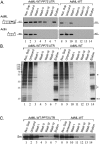
Analysis of affinity purified splicing complexes assembled on AdML-WT-PP73'UTR. Nuclear supernatants prepared from cell lines stably expressing the AdML-WT-PP73'UTR transcript, harboring the PP7 tag at the 3'UTR (left), or AdML-WT transcript without the tag (right), were affinity purified. Aliquots from the different steps of the affinity purification procedure were analyzed by RT-PCR (A); silver staining of SDS-PAGE (B); and WB (C). Lanes 1 and 8, Nuclear Supernatant (Nuc. Sup., 10%); Lanes 2 and 9, material not bound to the beads (Unbound); Lanes 3–6 and 10–13, washes; Lanes 7 and 14, elution. (A) RT-PCR products were electrophoresed on 1% agarose gel. Their identity is given on the left, open boxes represent exons, arrowheads represent the PCR primers. The expected size of each PCR product (nt) is given adjacent to its location on the left; (B) Proteins from the different steps of the affinity purification procedure were electrophoresed on 10% polyacrylamide gel and the gel was silver stained. Protein size marker is given on the left. *, heavy and light chains of the IgG antibody used for the affinity purification procedure; **, PP7CP protein; (C) Proteins from the different steps of the affinity purification procedure were electrophoresed on 12% polyacrylamide gel, transferred to a Nitrocellulose membrane and analyzed by WB using an anti-Sm antibody.
Figure 3
Figure 3
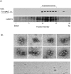
The affinity purified splicing complexes assembled in vivo on AdML-WT-PP73'UTR transcripts are supraspliceosomes. (A) Upper panel: Affinity purified splicing complexes assembled on the AdML-WT-PP73'UTR transcript, were fractionated in 10%–45% glycerol gradients, under the conditions used for fractionation of supraspliceosomes [15]. RNA was extracted from each fraction, analyzed by RT-PCR using the indicated AdML primer pair (arrowheads), and the amplified products were electrophoresed on 1% agarose gel. The size of the PCR product (nt) is given adjacent to its location, open boxes represent exons; Lower panel, Western Blot analysis of nuclear supernatants enriched for supraspliceosomes prepared from HeLa cells and fractionated in 10%–45% glycerol gradients. Aliquots from gradient fractions were analyzed by WB using anti-hnRNP G antibody. hnRNP G was previously shown to predominantly sediment with supraspliceosomes and to be associated with them [17]; (B) Upper panel, Gallery of electron micrographs of affinity purified supraspliceosomes assembled on AdML-WT-PP73'UTR transcript, visualized by EM, after negative staining with 1% uranyl acetate; Lower panel, Control, electron micrograph of eluted material from the control sample (AdML-WT transcript without the PP7 tag). The bar represents 50 nm.
Figure 4
Figure 4
Analysis of affinity purified suprasliceosomes assembled on AdML-Mut-PP73'UTR. Nuclear supernatants prepared from cell lines stably expressing the AdML-Mut-PP73'UTR transcript, harboring the PP7 tag at the 3'UTR (left) or AdML-Mut transcript without the tag (right) were affinity purified. Aliquots from the different steps of the affinity purification procedure were analyzed by RT-PCR (A); silver staining of SDS-PAGE (B) and WB (C). Lanes 1 and 8, Nuclear Supernatant (Nuc. Sup., 10%); Lanes 2 and 9, material not bound to the beads (Unbound); Lanes 3–6 and 10–13, washes; Lanes 7 and 14, elution. (A) RT-PCR products were electrophoresed on 1% agarose gel. Their identity is given on the left, open boxes represent exons, line intron, arrowheads represent PCR primers. The expected size of each PCR product (nt) is given adjacent to its location on the left; (B) Proteins from the different steps of the affinity purification procedure were electrophoresed on 10% polyacrylamide gel and the gel was silver stained. Protein size marker is given on the left. *, heavy and light chains of the IgG antibody used for the affinity purification procedure. **, PP7CP protein; (C) Proteins from the different steps of the affinity purification procedure were electrophoresed on 12% polyacrylamide gel, transferred to a Nitrocellulose membrane and analyzed by WB using an anti-Sm antibody; (D) EM visualization. Upper panel, Gallery of electron micrographs of affinity purified supraspliceosomes assembled on AdML-Mut-PP73'UTR transcript, visualized by EM, after negative staining with 1% uranyl acetate; Lower panel, Control, electron micrograph of eluted material from the control sample (AdML-Mut transcript without the PP7 tag). The bar represents 50 nm.
Figure 5
Figure 5
Analysis of affinity purified suprasliceosomes assembled on AdML-Mut-PP7IVS. Nuclear supernatants prepared from cell lines stably expressing the AdML-Mut-PP7IVS transcript, harboring the PP7 tag at the intron (left) or AdML-Mut transcript without the tag (right) were affinity purified. Aliquots from the different steps of the affinity purification procedure were analyzed by RT-PCR (A); silver staining of SDS-PAGE (B) and WB (C). Lanes 1 and 8, Nuclear Supernatant (Nuc. Sup., 10%); Lanes 2 and 9, material not bound to the beads (Unbound); Lanes 3–6 and 10–13, washes; Lanes 7 and 14, elution. (A) RT-PCR products were electrophoresed on 1% agarose gel. Their identity is given on the left, open boxes represent exons, line intron, arrowheads represent PCR primers. The expected size of each PCR product (nt) is given adjacent to its location on the left; (B) Proteins from the different steps of the affinity purification procedure were electrophoresed on 10% polyacrylamide gel and the gel was silver stained. Protein size marker is given on the left. *, heavy and light chains of the IgG antibody used for the affinity purification procedure. **, PP7CP protein; (C) Proteins from the different steps of the affinity purification procedure were electrophoresed on 12% polyacrylamide gel, transferred to a Nitrocellulose membrane and analyzed by WB using an anti-Sm antibody; (D) EM visualization. Upper panel, Gallery of electron micrographs of affinity purified supraspliceosomes assembled on AdML-Mut-PP7IVS transcript, visualized by EM, after negative staining with 1% uranyl acetate. Lower panel, Control, electron micrograph of eluted material from the control sample (AdML-Mut transcript without the PP7 tag). The bar represents 50 nm.
Figure 6
Figure 6
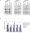
The five spliceosomal U snRNPs are associated with supraspliceosomes assembled in vivo at all splicing stages. (A) Northern blot analysis with probes directed against the five spliceosomal U snRNAs was performed on RNA extracted from nuclear supernatants and from affinity purified splicing complexes prepared from HeLa cell-lines expressing the AdML constructs either with the PP7 tag or without it (−), as indicated. Left, the AdML-WT-PP73'UTR transcript; middle, the AdML-Mut-PP73'UTR transcript; Right, the AdML-Mut-PP7IVS transcript. RNA was extracted either from the nuclear supernatant (Nuc. Sup.), or from the affinity purified samples (Elution). Aliquots were electrophoresed on 7 M urea/10% polyacrylamide gel, transferred to a nylon membrane, and hybridized with [α-32P] UTP-labeled RNA probes against the U1, U2, U4, U5, and U6 snRNAs, as described [15,16]. The identity of the U snRNA probes is given on the left; (B) Quantification of the Northern analyses. For each U snRNA molecule, the ratio between the signal of the eluted material and the relevant nuclear supernatant is shown. Each bar represents an average of two experiments; standard deviation is shown on the graph.
Figure 7
Figure 7
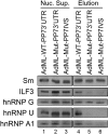
WB analyses of proteins associated with supraspliceosomes assembled on AdML-WT and AdML-Mut transcripts confirm the MS results. Nuclear supernatants were prepared from each of the HeLa cell lines, stably expressing either the AdML-WT-PP73'UTR, the AdML-Mut-PP73'UTR, or the AdML-Mut-PP7IVS transcripts, and supraspliceosomes assembled on each of the respective PP7-tagged transcripts were affinity purified, as described above. Proteins from each of the nuclear supernatants (Nuc. Sup., left) and from each the affinity purified supraspliceosomes (Elution, right) were extracted, electrophoresed on 8% polyacrylamide gel and analyzed by WB. The different antibodies used for the WB analyses are given on left.

Similar articles

Cited by

References

    1. Will C.L., Luhrmann R. Spliceosome structure and function. Cold Spring Harb. Perspect. Biol. 2011;3:a003707. - PMC - PubMed
    1. Burge C.B., Tuschl T.H., Sharp P.A. In: The RNA World. 2nd ed. Gesteland R.F., Cech T.R., Atkins J.F., editors. Cold Spring Harbor Laboratory Press; New York, NY, USA: 1999. pp. 525–560.
    1. Brow D.A. Allosteric cascade of spliceosome activation. Annu. Rev. Genet. 2002;36:333–360. - PubMed
    1. Will C.L., Lührmann R. Spliceosomal UsnRNP biogenesis, structure and function. Curr. Opin. Cell Biol. 2001;13:290–301. - PubMed
    1. Staley J.P., Guthrie C. Mechanical devices of the spliceosome: Motors, clocks, springs, and things. Cell. 1998;92:315–326. doi: 10.1016/S0092-8674(00)80925-3. - DOI - PubMed

Publication types

LinkOut - more resources