

doi: 10.1021/ja504845f.
Epub 2014 Jul 1.
Liposomal spherical nucleic acids
Affiliations
- PMID: 24983505
- PMCID: PMC4280063
- DOI: 10.1021/ja504845f
Item in Clipboard
Liposomal spherical nucleic acids
J Am Chem Soc.
.
Abstract
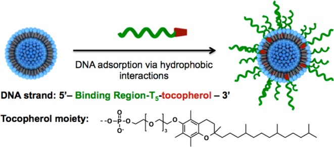
A novel class of metal-free spherical nucleic acid nanostructures was synthesized from readily available starting components. These particles consist of 30 nm liposomal cores, composed of an FDA-approved 1,2-dioleoyl-sn-glycero-3-phosphocholine (DOPC) lipid monomer. The surface of the liposomes was functionalized with DNA strands modified with a tocopherol tail that intercalates into the phospholipid layer of the liposomal core via hydrophobic interactions. The spherical nucleic acid architecture not only stabilizes these constructs but also facilitates cellular internalization and gene regulation in SKOV-3 cells.
Figures
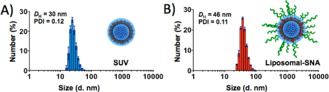
(A) DLS of SUVs after purification. (B) DLS of liposomal
SNAs after
purification.
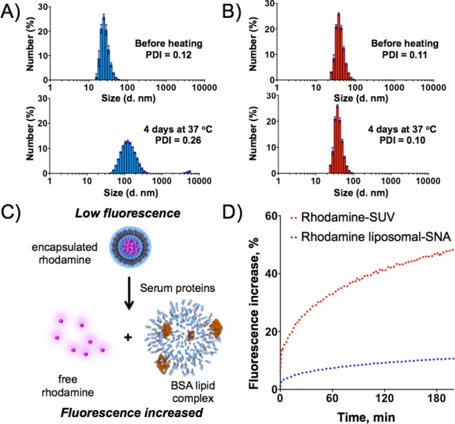
Stability studies of
SUV and liposomal SNAs. Change in average
diameter of SUVs (A) and liposomal SNAs (B) before (top) and after
(bottom) heating in buffer, as measured by DLS. (C) Schematic representation
of the decomposition of rhodamine-encapsulated liposome in the presence
of bovine serum albumin, a major component of FBS. (D) Degradation
of SUVs (red traces) and liposomal SNAs (blue traces) in the presence
of 10% FBS, as measured by increases in the fluorescence intensity
of rhodamine.
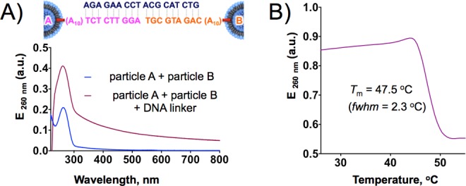
(A) Extinction spectra of liposomal SNAs before (blue) and after
(red) aggregation in the presence of linker DNA strands. (B) Melting
transition of liposomal SNA aggregates monitored as a change in extinction
at 260 nm.
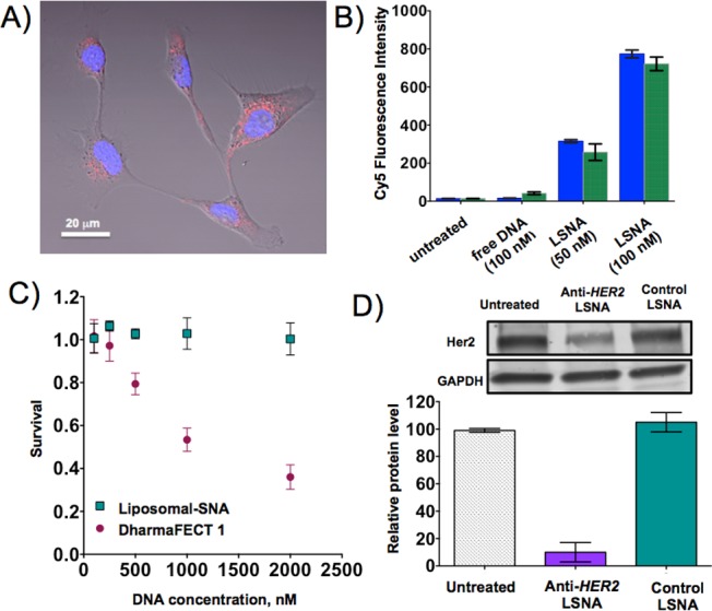
(A) Confocal micrograph of SKOV-3 cells
incubated with 100 nM Cy5-labeled
liposomal SNAs (red) for 24 h. (B) Flow cytometry analysis of uptake
of 5-Cy5-labeled DNA strand and 5′-Cy5-labeled liposomal SNAs
in SKOV-3 cells after 1 h (blue bars) and 36 h (green bars) of incubation.
(C) Cytotoxicity (alamarBlue assay) of liposomal SNAs and DharmaFECT-delivered
DNA in SKOV-3 cells. (D) HER2 gene knockdown in SKOV-3
cells using anti-HER2 liposomal SNA constructs at
1 μM DNA.
References
-
- Mirkin C. A.; Letsinger R. L.; Mucic R. C.; Storhoff J. J. Nature 1996, 382, 607. - PubMed
-
- Cutler J. I.; Auyeung E.; Mirkin C. A. J. Am. Chem. Soc. 2012, 134, 1376. - PubMed
- Briley W.; Halo T. L.; Randeria P. S.; Alhasan A. H.; Auyeung E.; Hurst S. J.; Mirkin C. A. In Nanomaterials for Biomedicine; Nagarajan R., Ed.; ACS Symposium Series1119; American Chemical Society: Washington, DC, 2012; pp 1–20.
Publication types
MeSH terms
Substances
Grants and funding
LinkOut - more resources
Full Text Sources
Other Literature Sources
