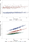
Variation in the percent of emphysema-like lung in a healthy, nonsmoking multiethnic sample. The MESA lung study
- PMID: 24983825
- PMCID: PMC4213995
- DOI: 10.1513/AnnalsATS.201310-364OC
Variation in the percent of emphysema-like lung in a healthy, nonsmoking multiethnic sample. The MESA lung study
Abstract
Rationale: Computed tomography (CT)-based lung density is used to quantitate the percentage of emphysema-like lung (hereafter referred to as percent emphysema), but information on its distribution among healthy nonsmokers is limited.
Objectives: We evaluated percent emphysema and total lung volume on CT scans of healthy never-smokers in a multiethnic, population-based study.
Methods: The Multi-Ethnic Study of Atherosclerosis (MESA) Lung Study investigators acquired full-lung CT scans of 3,137 participants (ages 54-93 yr) between 2010-12. The CT scans were taken at full inspiration following the Subpopulations and Intermediate Outcome Measures in COPD Study (SPIROMICS) protocol. "Healthy never-smokers" were defined as participants without a history of tobacco smoking or respiratory symptoms and disease. "Percent emphysema" was defined as the percentage of lung voxels below -950 Hounsfield units. "Total lung volume" was defined by the volume of lung voxels.
Measurements and main results: Among 854 healthy never-smokers, the median percent emphysema visualized on full-lung scans was 1.1% (interquartile range, 0.5-2.5%). The percent emphysema values were 1.2 percentage points higher among men compared with women and 0.7, 1.2, and 1.2 percentage points lower among African Americans, Hispanics, and Asians compared with whites, respectively (P < 0.001). Percent emphysema was positively related to age and height and inversely related to body mass index. The findings were similar for total lung volume on CT scans and for percent emphysema defined at -910 Hounsfield units and measured on cardiac scans. Reference equations to account for these differences are presented for never, former and current smokers.
Conclusions: Similar to lung function, percent emphysema varies substantially by demographic factors and body size among healthy never-smokers. The presented reference equations will assist in defining abnormal values for percent emphysema and total lung volume on CT scans, although validation is pending.
Keywords: emphysema; lung volumes; quantitative computed tomography; reference equations.
Figures


References
-
- Hoffman EA. Effect of body orientation on regional lung expansion: a computed tomographic approach. J Appl Physiol (1985) 1985;59:468–480. - PubMed
-
- Coxson HO, Mayo JR, Behzad H, Moore BJ, Verburgt LM, Staples CA, Paré PD, Hogg JC. Measurement of lung expansion with computed tomography and comparison with quantitative histology. J Appl Physiol (1985) 1995;79:1525–1530. - PubMed
-
- Gevenois PA, de Maertelaer V, De Vuyst P, Zanen J, Yernault JC. Comparison of computed density and macroscopic morphometry in pulmonary emphysema. Am J Respir Crit Care Med. 1995;152:653–657. - PubMed
Publication types
MeSH terms
Grants and funding
- N01 HC95163/HC/NHLBI NIH HHS/United States
- N01 HC95165/HC/NHLBI NIH HHS/United States
- N01 HC95168/HC/NHLBI NIH HHS/United States
- N01 HC95164/HC/NHLBI NIH HHS/United States
- R01 HL077612/HL/NHLBI NIH HHS/United States
- HL064368/HL/NHLBI NIH HHS/United States
- HL112986/HL/NHLBI NIH HHS/United States
- N01 HC95169/HC/NHLBI NIH HHS/United States
- N01 HC95162/HC/NHLBI NIH HHS/United States
- N01 HC95167/HC/NHLBI NIH HHS/United States
- R01 HL064368/HL/NHLBI NIH HHS/United States
- P30 ES009089/ES/NIEHS NIH HHS/United States
- R01 HL112986/HL/NHLBI NIH HHS/United States
- N01 HC095169/HL/NHLBI NIH HHS/United States
- R01 HL093081/HL/NHLBI NIH HHS/United States
- N01 HC95159/HC/NHLBI NIH HHS/United States
- N01 HC95160/HC/NHLBI NIH HHS/United States
- N01 HC95161/HC/NHLBI NIH HHS/United States
- N01 HC95166/HC/NHLBI NIH HHS/United States
LinkOut - more resources
Full Text Sources
Other Literature Sources

