Immune-directed support of rich microbial communities in the gut has ancient roots
- PMID: 24984114
- PMCID: PMC4146740
- DOI: 10.1016/j.dci.2014.06.011
Immune-directed support of rich microbial communities in the gut has ancient roots
Abstract
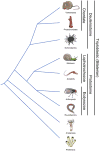
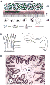
The animal gut serves as a primary location for the complex host-microbe interplay that is essential for homeostasis and may also reflect the types of ancient selective pressures that spawned the emergence of immunity in metazoans. In this review, we present a phylogenetic survey of gut host-microbe interactions and suggest that host defense systems arose not only to protect tissue directly from pathogenic attack but also to actively support growth of specific communities of mutualists. This functional dichotomy resulted in the evolution of immune systems much more tuned for harmonious existence with microbes than previously thought, existing as dynamic but primarily cooperative entities in the present day. We further present the protochordate Ciona intestinalis as a promising model for studying gut host-bacterial dialogue. The taxonomic position, gut physiology and experimental tractability of Ciona offer unique advantages in dissecting host-microbe interplay and can complement studies in other model systems.
Keywords: Ciona intestinalis; Gut biofilms; Gut host–microbe interactions; Gut immune effectors; Gut immune evolution; Mutualism.
Copyright © 2014 Elsevier Ltd. All rights reserved.
Figures


Similar articles
-
Reflections on the Use of an Invertebrate Chordate Model System for Studies of Gut Microbial Immune Interactions.Front Immunol. 2021 Feb 25;12:642687. doi: 10.3389/fimmu.2021.642687. eCollection 2021. Front Immunol. 2021. PMID: 33717199 Free PMC article. Review.
-
The gut of geographically disparate Ciona intestinalis harbors a core microbiota.PLoS One. 2014 Apr 2;9(4):e93386. doi: 10.1371/journal.pone.0093386. eCollection 2014. PLoS One. 2014. PMID: 24695540 Free PMC article.
-
The gut virome of the protochordate model organism, Ciona intestinalis subtype A.Virus Res. 2018 Jan 15;244:137-146. doi: 10.1016/j.virusres.2017.11.015. Epub 2017 Nov 15. Virus Res. 2018. PMID: 29155033
-
An Immune Effector System in the Protochordate Gut Sheds Light on Fundamental Aspects of Vertebrate Immunity.Results Probl Cell Differ. 2015;57:159-73. doi: 10.1007/978-3-319-20819-0_7. Results Probl Cell Differ. 2015. PMID: 26537381 Review.
-
Generation of Germ-Free Ciona intestinalis for Studies of Gut-Microbe Interactions.Front Microbiol. 2016 Dec 27;7:2092. doi: 10.3389/fmicb.2016.02092. eCollection 2016. Front Microbiol. 2016. PMID: 28082961 Free PMC article.
Cited by
-
Environmental stress and nanoplastics' effects on Ciona robusta: regulation of immune/stress-related genes and induction of innate memory in pharynx and gut.Front Immunol. 2023 May 29;14:1176982. doi: 10.3389/fimmu.2023.1176982. eCollection 2023. Front Immunol. 2023. PMID: 37313415 Free PMC article.
-
Gut Microbiomes of the Eastern Oyster (Crassostrea virginica) and the Blue Mussel (Mytilus edulis): Temporal Variation and the Influence of Marine Aggregate-Associated Microbial Communities.mSphere. 2019 Dec 11;4(6):e00730-19. doi: 10.1128/mSphere.00730-19. mSphere. 2019. PMID: 31826972 Free PMC article.
-
Markedly Elevated Antibody Responses in Wild versus Captive Spotted Hyenas Show that Environmental and Ecological Factors Are Important Modulators of Immunity.PLoS One. 2015 Oct 7;10(10):e0137679. doi: 10.1371/journal.pone.0137679. eCollection 2015. PLoS One. 2015. PMID: 26444876 Free PMC article.
-
Immunity in Protochordates: The Tunicate Perspective.Front Immunol. 2017 Jun 9;8:674. doi: 10.3389/fimmu.2017.00674. eCollection 2017. Front Immunol. 2017. PMID: 28649250 Free PMC article. Review.
-
Development of the preterm infant gut microbiome: a research priority.Microbiome. 2014 Oct 13;2:38. doi: 10.1186/2049-2618-2-38. eCollection 2014. Microbiome. 2014. PMID: 25332768 Free PMC article. Review.
References
-
- Abreu MT. Toll-like receptor signalling in the intestinal epithelium: how bacterial recognition shapes intestinal function. Nat Rev Immunol. 2010;10:131–144. - PubMed
-
- Agostini S, Suzuki Y, Higuchi T, Casareto BE, Yoshinaga K, Nakano Y, Fujimura H. Biological and chemical characteristics of the coral gastric cavity. Coral Reefs. 2012;31:147–156.
-
- Amaro T, Witte H, Herndl GJ, Cunha MR, Billett DSM. Deep-sea bacterial communities in sediments and guts of deposit-feeding holothurians in Portuguese canyons (NE Atlantic) Deep-Sea Research I. 2009;56:1834–1843.
-
- Artis D. Epithelial-cell recognition of commensal bacteria and maintenance of immune homeostasis in the gut. Nat Rev Immunol. 2008;8:411–420. - PubMed
Publication types
MeSH terms
Grants and funding
LinkOut - more resources
Full Text Sources
Other Literature Sources

