. 2014 Jul 1:68:1.5.1-1.5.39.
doi: 10.1002/0471142301.ns0105s68.

Transneuronal circuit analysis with pseudorabies viruses

Affiliations

Transneuronal circuit analysis with pseudorabies viruses

J Patrick Card et al. Curr Protoc Neurosci. .

Abstract

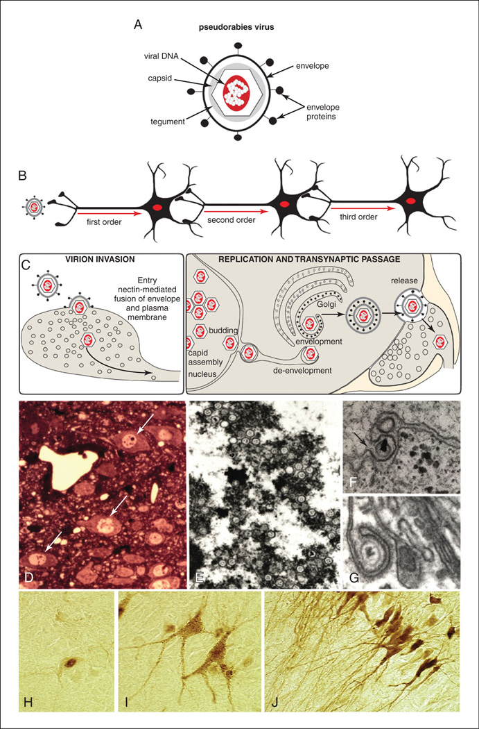
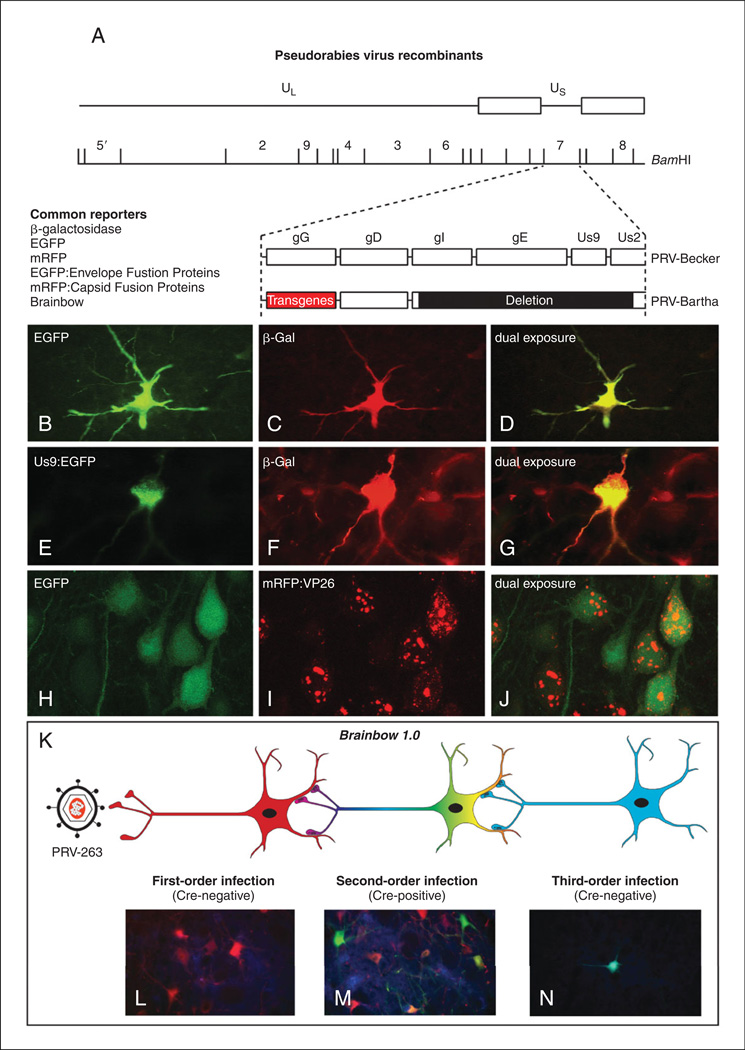
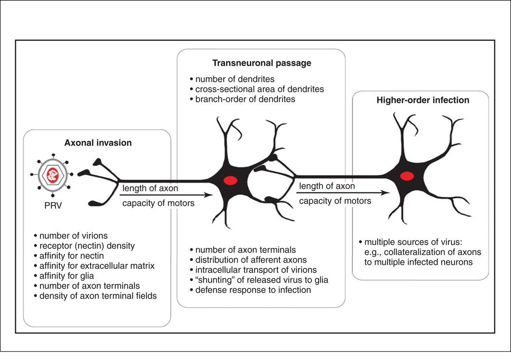
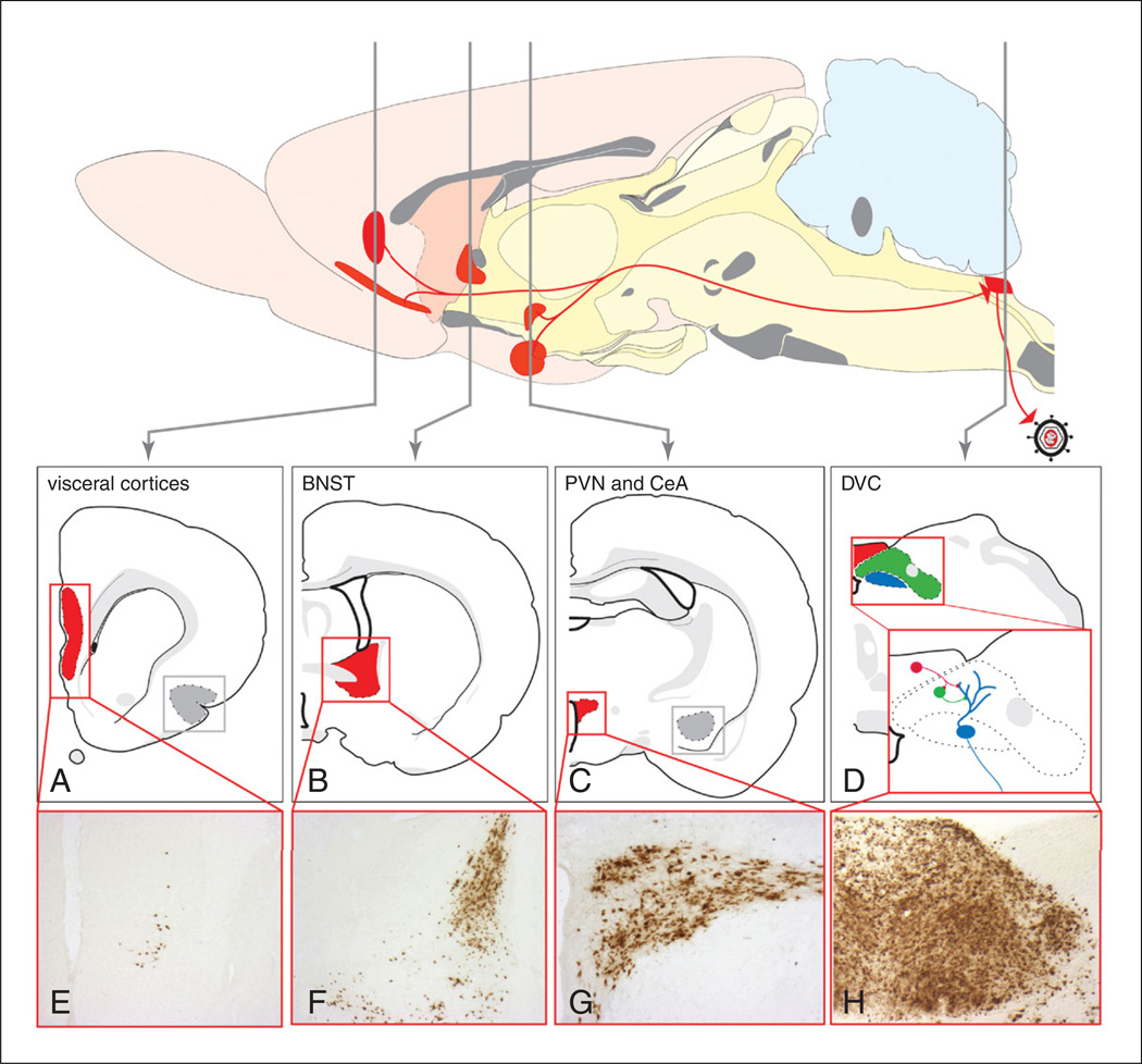
Our ability to understand the function of the nervous system is dependent upon defining the connections of its constituent neurons. Development of methods to define connections within neural networks has always been a growth industry in the neurosciences. Transneuronal spread of neurotropic viruses currently represents the best means of defining synaptic connections within neural networks. The method exploits the ability of viruses to invade neurons, replicate, and spread through the intimate synaptic connections that enable communication among neurons. Since the method was first introduced in the 1970s, it has benefited from an increased understanding of the virus life cycle, the function of viral genome, and the ability to manipulate the viral genome in support of directional spread of virus and the expression of transgenes. In this unit, we review these advances in viral tracing technology and the way in which they may be applied for functional dissection of neural networks.

Keywords: herpesvirus; rabies; transgene expression; transneuronal.


Figures

Figure 1.5.1
Figure 1.5.1
(A) The structure of alpha herpesvirus virions and features characteristic of their neuroinvasiveness are illustrated. Viral DNA is sequestered within a capsid composed of virally encoded proteins. The capsid and a surrounding tegument of each virion are contained within a viral envelope acquired from the host cell. The envelope contains a second set of virally encoded proteins that are important for target cell recognition, attachment, and the receptor-mediated fusion event that leads to the release of the capsid into a permissive cell. (B) Recombinant strains of PRV-Bartha move selectively in the retrograde direction through neural circuits. (C) A model of virion assembly postulated for pseudorabies virus is illustrated. Assembly of virions is a multistep process that leads to assembly of mature virions in the cell soma. Virions traffic through the soma and dendrites of infected cells and are released in the vicinity of synaptic contacts. Adapted with permission from Card (1998). (D) Distinct nuclear inclusions (arrows) mark infected neurons and are clinically diagnostic for herpesvirus infection. (E) Transmission electron microscopy reveals that nuclear inclusions are sites of capsid assembly. (F) Capsids bud through the inner leaf of the nuclear envelope (arrow) in transit to the cytoplasm, where they acquire lipid bylayers from the trans-Golgi reticulum (G) or late endosomal compartment. (H-J) The distribution of viral antigens within infected cells provides an index of the stage of infection. At early stages (H), antigens are largely confined to the cell nucleus. As capsids migrate into the cell cytoplasm and acquire an envelope, viral antigens appear throughout the soma and proximal dendrites (I). Punctate staining on the cell soma and dendrites marks sites of transneuronal passage. At late stages of infection (J), viral antigens fill the entirety of the dendritic compartment.
Figure 1.5.2
Figure 1.5.2
(A) The organization of the PRV-Bartha genome and a common site of transgene insertion (gG) for recombinant viruses is illustrated. (B-J) Reporter expression from recombinant viruses useful in single- and dual-infection studies is illustrated. (C-D) In this case, either bacterial β-galactosidase (β-Gal) or jellyfish enhanced green fluorescent protein (EGFP) genes have been engineered into the gG locus, and the cell was infected by retrograde transport from two different projection targets from a collateralized axon. (E-G) Dual infection in the same paradigm resulting from replication of β-Gal and an EGFP Us9 fusion protein is illustrated. Note the differential concentration of the fusion protein in the Golgi complex of the cell cytoplasm. (H-I) Dual infection of cells with HSV-129 recombinant viruses expressing EGFP and a mRVP-capsid (VP26) fusion protein is illustrated. The fusion protein is differentially concentrated in the cell nucleus, whereas EGFP is a cytoplasmic marker, making dual-infected cells easily identified. (K) The experimental paradigm employing Cre-Lox technology for conditional expression of reporters from the Brainbow 1.0 cassette is illustrated. (L) The dTomato (red) reporter is the default maker of infected cells in the absence of Cre. (M) In the presence of Cre, the dTomato reporter is recombined from the viral genome to enable the expression of EYFP and mCerulean (cyan) reporters. (N) Neurons infected by transneuronal passage of recombined virus will only express the conditional reporters.
Figure 1.5.3
Figure 1.5.3
The factors that influence replication and spread of PRV-Bartha recombinants through neural circuits are illustrated.
Figure 1.5.4
Figure 1.5.4
The sequential retrograde trans-synaptic passage of PRV-Bartha from the stomach wall of the rat is illustrated. Retrograde transport of virus from the stomach wall results in infection of parasympathetic neurons in the dorsal motor vagal nucleus (blue in D) and transneuronal spread of virus to neurons in the adjacent NTS (green in D) and area postrema (red in D). Replication and spread of infection to higher-order neurons occurs with advancing survival. Included in this extended network is the paraventricular hypothalamic nucleus (C and G; PVN), central nucleus of the amygdala (CeA), bed nucleus of the stria terminalis (B,F; BNST), and visceral cortices (A,E). This model has been used extensively to define the synaptic organization of preautonomic neural networks and to define approaches for statistical evaluation of the progression of infection. See Card et al. (2005a) for details.

References

    1. Archin NC, van den Boom L, Perelygina L, Hilliard JM, Atherton SS. Delayed spread and reduction in virus titer after anterior chamber inoculation of a recombinant of HSV-1 expressing IL-16. Invest. Ophthalmol. Vis. Sci. 2003;44:3066–3076. - PubMed
    1. Archin NM, Atherton SS. Rapid spread of a neurovirulent strain of HSV-1 through the CNS of BALB/c mice following anterior chamber inoculation. J. Neurovirol. 2002;8:122–135. - PubMed
    1. Bamshad M, Aoki VT, Adkison MG, Warren WS, Bartness TJ. Central nervous system origins of the sympathetic nervous system out flow to white adipose tissue. Am. J. Physiol. 1998;275:R291–R299. - PubMed
    1. Bamshad M, Song CK, Bartness TJ. CNS origins of the sympathetic nervous system outflow to brown adipose tissue. Am. J. Physiol. 1999;45:R1569–R1578. - PubMed
    1. Banfield BW, Kaufman JD, Randall JA, Pickard GE. Development of pseudorabies virus strains expressing red fluorescent proteins: New tools for multisynaptic labeling applications. J. Virol. 2003;77:10106–10112. - PMC - PubMed

Publication types

LinkOut - more resources