An official website of the United States government
The .gov means it’s official.
Federal government websites often end in .gov or .mil. Before
sharing sensitive information, make sure you’re on a federal
government site.
The site is secure.
The https:// ensures that you are connecting to the
official website and that any information you provide is encrypted
and transmitted securely.
A critical requirement for mitosis is the distribution of genetic material to the two daughter cells. The central player in this process is the macromolecular kinetochore structure, which binds to both chromosomal DNA and spindle microtubule polymers to direct chromosome alignment and segregation. This review will discuss the key kinetochore activities required for mitotic chromosome segregation, including the recognition of a specific site on each chromosome, kinetochore assembly and the formation of kinetochore-microtubule connections, the generation of force to drive chromosome segregation, and the regulation of kinetochore function to ensure that chromosome segregation occurs with high fidelity.
Core requirements for DNA segregation. Cartoon diagram showing the core activities required for…
Figure 1.
Core requirements for DNA segregation. Cartoon diagram showing the core activities required for DNA segregation of the bacterial R1 plasmid or eukaryotic chromosomes highlighting the recognition of DNA, physical connections, and force.
Figure 2.
Recognition. ( A ) Diagram…
Figure 2.
Recognition. ( A ) Diagram showing the key players and processes required for…
Figure 2.
Recognition. (A) Diagram showing the key players and processes required for the deposition of the specialized CENP-A-containing nucleosomes at centromeres. (B) (Top) Diagram of the key components of centromeric chromatin. (Bottom) Crystal structures of the H3/H4 tetramer based on data from Davey et al. (2002), CENP-A/H4 tetramer based on data from Tachiwana et al. (2011), and CENP-T/W/S/X heterotetramer based on data from Nishino et al. (2012).
Attachment. Diagram showing kinetochore structure and organization during interphase and mitosis. At mitotic…
Figure 3.
Attachment. Diagram showing kinetochore structure and organization during interphase and mitosis. At mitotic entry, CDK/Cyclin B phosphorylation promotes outer kinetochore assembly on a platform of constitutive kinetochore proteins. For information on the additional kinetochore proteins shown in the figure, see Cheeseman and Desai (2008).
Figure 4.
Force. Diagrams showing the kinetochore…
Figure 4.
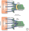
Force. Diagrams showing the kinetochore associating with depolymerizing microtubules and harnessing the force…
Figure 4.
Force. Diagrams showing the kinetochore associating with depolymerizing microtubules and harnessing the force induced by microtubule depolymerization to direct chromosome movement. The Ndc80 complex is the core player in forming kinetochore–microtubule interactions, but it requires additional interactions with the Dam1 complex (fungi, top) or the Ska1 complex (vertebrates, bottom).
Figure 5.
Regulation. ( A ) Diagram…
Figure 5.
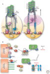
Regulation. ( A ) Diagram showing the tension-dependent deformation of the kinetochore structure.…
Figure 5.
Regulation. (A) Diagram showing the tension-dependent deformation of the kinetochore structure. Aurora B kinase located at the inner centromere (at the base of the kinetochore) is spatially separated from its substrates at the outer kinetochore. The presence of tension on bi-oriented kinetochores strongly reduces the ability of Aurora B to phosphorylate outer kinetochore proteins and inactivate microtubule attachments. (B) Model showing the spindle assembly checkpoint proteins preventing cell cycle progression in the absence of unattached kinetochores.
Al-Bassam J, Kim H, Brouhard G, van Oijen A, Harrison SC, Chang F 2010. CLASP promotes microtubule rescue by recruiting tubulin dimers to the microtubule. Dev Cell 19: 245–258
-
PMC
-
PubMed
Al-Bassam J, Chang F 2011. Regulation of microtubule dynamics by TOG-domain proteins XMAP215/Dis1 and CLASP. Trends Cell Biol 21: 604–614
-
PMC
-
PubMed
Amor DJ, Bentley K, Ryan J, Perry J, Wong L, Slater H, Choo KH 2004. Human centromere repositioning “in progress”. Proc Natl Acad Sci 101: 6542–6547
-
PMC
-
PubMed
Asbury CL, Gestaut DR, Powers AF, Franck AD, Davis TN 2006. The Dam1 kinetochore complex harnesses microtubule dynamics to produce force and movement. Proc Natl Acad Sci 103: 9873–9878
-
PMC
-
PubMed
Bader JR, Vaughan KT 2010. Dynein at the kinetochore: Timing, interactions and functions. Semin Cell Dev Biol 21: 269–275
-
PMC
-
PubMed