Review
. 2014 Jul 1;6(7):a018762.
doi: 10.1101/cshperspect.a018762.

Writers and readers of histone acetylation: structure, mechanism, and inhibition

Affiliations
Review

Writers and readers of histone acetylation: structure, mechanism, and inhibition

Ronen Marmorstein et al. Cold Spring Harb Perspect Biol. .

Abstract

Histone acetylation marks are written by histone acetyltransferases (HATs) and read by bromodomains (BrDs), and less commonly by other protein modules. These proteins regulate many transcription-mediated biological processes, and their aberrant activities are correlated with several human diseases. Consequently, small molecule HAT and BrD inhibitors with therapeutic potential have been developed. Structural and biochemical studies of HATs and BrDs have revealed that HATs fall into distinct subfamilies containing a structurally related core for cofactor binding, but divergent flanking regions for substrate-specific binding, catalysis, and autoregulation. BrDs adopt a conserved left-handed four-helix bundle to recognize acetyllysine; divergent loop residues contribute to substrate-specific acetyllysine recognition.


Figures

Figure 1.
Figure 1.
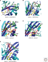
Overall structure of HAT proteins. Representative members of the five HAT subfamilies are illustrated as cartoons highlighting the structurally conserved core region (blue) and flanking amino- and carboxy-terminal regions (aqua). The cofactor is shown in stick figure in CPK coloring (carbon, yellow; oxygen, red; nitrogen, blue; phosphorous, orange; sulfur, brown): (A) yeast HAT1/AcCoA (PDB 1BOB), (B) Tetrahymena Gcn5/CoA/histone H3 (PDB code: 1PUA) with the histone H3 peptide shown in red, (C) yeast Esa1/H4K16-CoA (PDB code: 3TO6), (D) human p300/Lys-CoA (PDB code: 3BIY) with the substrate-binding loop shown in red, (E) yeast Rtt109/CoA (PDB code: 3D35) with the substrate-binding loop shown in red.
Figure 2.
Figure 2.
Catalytic mechanism of HAT proteins. Active sites of representative members of the HAT subfamilies are illustrated highlighting the relevant side chains on a backbone cartoon of the active site. (A) Tetrahymena Gcn5/CoA/histone H3. Key catalytic residues are labeled and hydrophobic residues of the active site that likely raise the pKa of Glu 173 are shown in stick figure in CPK coloring with carbon in green. A segment of the histone H3 peptide is shown in red. W indicates a well-ordered water molecule that participates in catalysis. The numbering is for yeast Gcn5. (B) Yeast Esa1 bound to the H4K16CoA bisubstrate inhibitor (stick figure and CPK coloring with carbon atoms in yellow). Key catalytic residues are labeled and hydrophobic residues of the active site that likely raise the pKa of Glu 338 are shown. Residues flanking K16 in the peptide are disordered in the structure. (C) Human p300 bound to the Lys-CoA bisubstrate inhibitor (stick figure and CPK coloring with carbon atoms in yellow). Residues shown to play catalytic roles are labeled with other potential catalytic residues shown in stick figure. The substrate-binding loop is shown in red. (D) Yeast Rtt109/CoA. Potential catalytic residues in the corresponding position of hp300 are shown. The CoA molecule is shown in stick figure in CPK coloring with carbon atoms in yellow. The substrate-binding loop is shown in red. (E) hHAT1/AcCoA/histone H4. The three general base candidate residues are represented as green stick figures and a segment of the histone H4 peptide is shown in red.
Figure 3.
Figure 3.
Histone substrate binding by HAT proteins. Close-up electrostatic view of HAT domain structures with histone peptide substrates or CoA-peptide bisubstrate inhibitors. Protein surfaces are colored according to electrostatic potential with the degree of red, blue, and white coloring correlating with electronegative, electropositive, and neutral charge, respectively. (A) Structure of tGcn5 bound to CoA (CPK coloring with carbon atoms shown in yellow) and a 19-residue histone H3 peptide (CPK coloring with carbon atoms shown in purple) centered around K14. (B) Structure of hHAT1 bound to AcCoA (CPK coloring with carbon atoms shown in yellow) and a 20-residue histone H4 peptide (CPK coloring with carbon atoms shown in purple) centered around K12. (C) Structure of the hp300/LysCoA complex. The LysCoA bisubstrate inhibitor is shown in CPK coloring with carbon atoms in yellow. (D) Structure of the yEsa1/H4K16CoA complex (only the lysine side chain of the H4K16 peptide component of the bisubstrate inhibitor is ordered in the crystal structure and shown in CPK coloring with carbon atoms in yellow).
Figure 4.
Figure 4.
Autoacetylation regulation of HAT proteins. Close-up views of the autoacetylation site of HAT proteins. (A) Model for p300 activation by autoacetylation. The black loop and green acetylated lysine balls are modeled on the p300/Lys-CoA crystal structure. (B) Structure of the K290 autoacetylation site of Rtt109, highlighting the environment around acetylated K290. The acetylated lysine and other side chains that interact with the acetylated lysine are indicated in stick figure in CPK coloring with carbon in green and the hydrogen bond is shown as a dotted orange line. The AcCoA molecule is shown as a stick figure in CPK coloring with carbon atoms in yellow. (C) Structure of the yEsa1/H4K16CoA complex is shown, highlighting the environment around acetylated K262 (green). The corresponding K274 of hMOF is superimposed in the unacetylated (yellow) and acetylated (orange) conformations showing that the unacetylated conformation would clash with binding of the cognate substrate lysine (as represented by the lysine of the H4K16CoA bisubstrate inhibitor shown in purple).
Figure 5.
Figure 5.
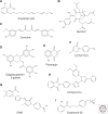
HAT inhibitors. Reported inhibitor specificities are as follows: (A,B,F) specific to p300 and PCAF; (CE) specific to p300; (G) specific to p300; (H,I) specific to MYST.
Figure 6.
Figure 6.
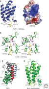
Bromodomains as acetyllysine-binding domains. In all structures, the histone peptide is in yellow and the main and side chains of the protein residues are color-coded by atom type. (A) The three-dimensional solution structure of the PCAF bromodomain bound to an H3K36ac peptide (PDB code: 2RNX) is illustrated as a ribbon diagram (left) and a surface electrostatic representation (right) of the protein with red and blue colors representing negatively or positively charged amino acid residues, respectively. (B) The acetyllysine-binding pocket is depicted from the crystal structure of the GCN5 bromodomain (green) in complex with an H4K16ac peptide (PDB code: 1E6I). This stick diagram shows key residues and bound water molecules (magenta spheres) contributing to acetyllysine recognition. Hydrogen bonding interactions are indicated by dotted lines. (C) The crystal structure of the tandem bromodomains of uncomplexed human TAF1 (PDB code: 1EQF). (D) The crystal structure of the first bromodomain of Brdt bound to an H4K5acK8ac peptide (PDB code: 2WP2).
Figure 7.
Figure 7.
The phylogenetic tree of human bromodomains. Sequence similarity–based dendrogram of the human bromodomains was generated using the neighbor-joining method with MEGA (Kumar et al. 2004). Sequences of the human bromodomains were obtained from the SMART database (Letunic et al. 2004) and aligned with SMART bromodomains’ hidden Markov models using Hmmalign (Sonnhammer et al. 1997). (Modified from Zhang et al. 2010.)
Figure 8.
Figure 8.
Interdomain interactions in tandem histone-binding modules. (A) The solution structure of the PHD (navy)–bromodomain (red and green) module of human KAP1 (PDB code: 2RO1). (B) The PHD–bromodomain module of human BPTF in complex with an H3K4 peptide (PDB code: 3QZV). (C) The crystal structure of the PHD–bromodomain module of human TRIM33 in complex with an H3K9me3K18acK23ac peptide (PDB code: 3U5P). Note that the second bromodomain in each of the above tandem modules are colored green, and each structure is oriented with respect to the αZ helix (red) of this bromodomain. (D) The solution structure of the tandem PHD finger module of human DPF3b bound to an H3K14ac peptide (PDB code: 2KWJ). The zinc atoms are highlighted as red spheres, and the main and side chains of the protein residues involved in H3K14ac binding are color-coded by atom type with green, red, and blue for carbon, oxygen and nitrogen, respectively.
Figure 9.
Figure 9.
Small molecule inhibitors of bromodomains. (A) Chemical structures of representative small-molecule bromodomain inhibitors, including NP1 (for PCAF bromodomain), ischemin, JQ1, I-BET and I-BET151. (B) Ischemin, a small-molecule inhibitor developed for the CBP bromodomain, depicted in a complexed 3D structure bound to the protein (PDB code: 2L84). (C) JQ1, a BET bromodomain-specific inhibitor, shown when bound to the first bromodomain of BRD4 in the crystal structure (PDB code: 3MXF).

References

    1. Ai X, Parthun MR 2004. The nuclear Hat1p/Hat2p complex: A molecular link between type B histone acetyltransferases and chromatin assembly. Mol Cell 14: 195–205 - PubMed
    1. Akella JS, Wloga D, Kim J, Starostina NG, Lyons-Abbott S, Morrissette NS, Dougan ST, Kipreos ET, Gaertig J 2010. MEC-17 is an α-tubulin acetyltransferase. Nature 467: 218–222 - PMC - PubMed
    1. Akhtar A, Becker PB 2001. The histone H4 acetyltransferase MOF uses a C2HC zinc finger for substrate recognition. EMBO Rep 2: 113–118 - PMC - PubMed
    1. Albaugh BN, Kolonko EM, Denu JM 2010. Kinetic mechanism of the Rtt109–Vps75 histone acetyltransferase-chaperone complex. Biochemistry 49: 6375–6385 - PMC - PubMed
    1. Albaugh BN, Arnold KM, Lee S, Denu JM 2011. Autoacetylation of the histone acetyltransferase RTT109. J Biol Chem 286: 24694–24701 - PMC - PubMed

Publication types

MeSH terms