

Attenuation of replication stress-induced premature cellular senescence to assess anti-aging modalities
- PMID: 24984966
- PMCID: PMC4156839
- DOI: 10.1002/0471142956.cy0947s69
Attenuation of replication stress-induced premature cellular senescence to assess anti-aging modalities
Abstract
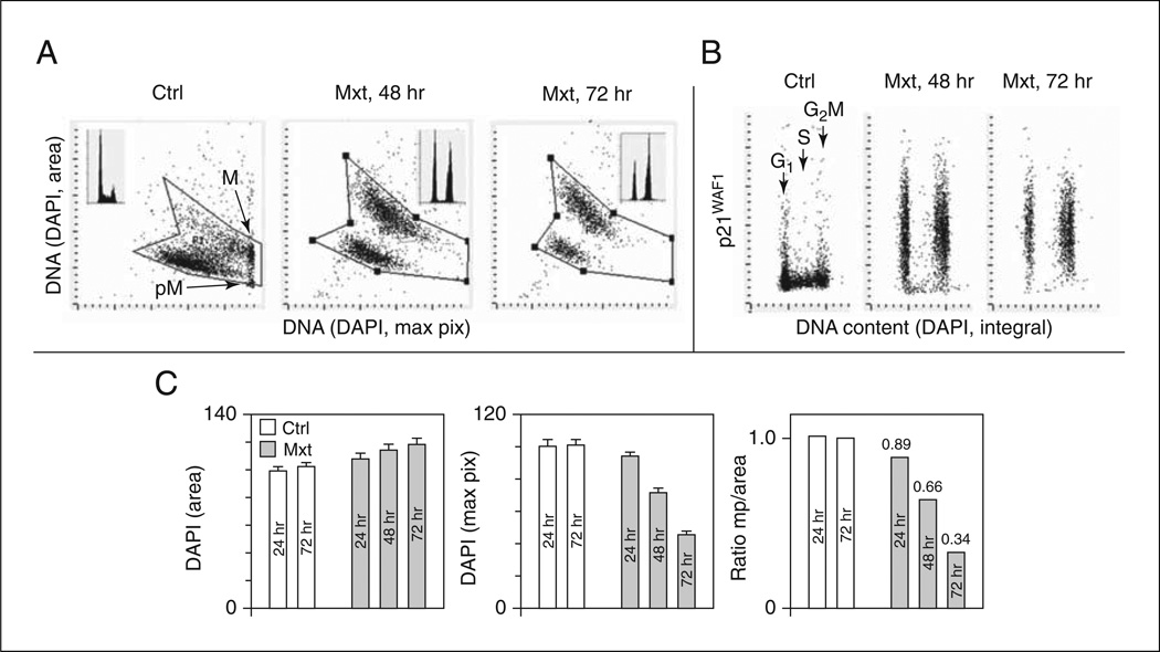
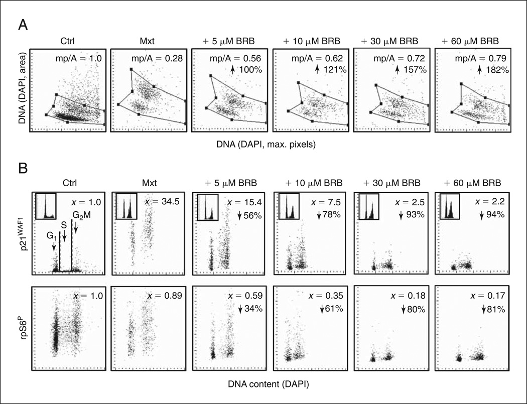
Described is an in vitro model of premature senescence in pulmonary adenocarcinoma A549 cells induced by persistent DNA replication stress in response to treatment with the DNA damaging drug mitoxantrone (Mxt). The degree of cellular senescence, based on characteristic changes in cell morphology, is measured by laser scanning cytometry. Specifically, the flattening of cells grown on slides (considered the hallmark of cellular senescence) is measured as the decline in local intensity of DNA-associated DAPI fluorescence (represented by maximal pixels). This change is paralleled by an increase in nuclear area. Thus, the ratio of mean intensity of maximal pixels to nuclear area provides a very sensitive morphometric biomarker for the degree of senescence. This analysis is combined with immunocytochemical detection of senescence markers, such as overexpression of cyclin kinase inhibitors (e.g., p21(WAF1) ) and phosphorylation of ribosomal protein S6 (rpS6), a key marker associated with aging/senescence that is detected using a phospho-specific antibody. These biomarker indices are presented in quantitative terms defined as a senescence index (SI), which is the fraction of the marker in test cultures relative to the same marker in exponentially growing control cultures. This system can be used to evaluate the anti-aging potential of test agents by assessing attenuation of maximal senescence. As an example, the inclusion of berberine, a natural alkaloid with reported anti-aging properties and a long history of use in traditional Chinese medicine, is shown to markedly attenuate the Mxt-induced SI and phosphorylation of rpS6. The multivariate analysis of senescence markers by laser scanning cytometry offers a promising tool to explore the potential anti-aging properties of a variety agents.
Keywords: berberine; cell cycle; laser scanning cytometry; mTOR signaling; nuclear size; ribosomal protein S6.
Copyright © 2014 John Wiley & Sons, Inc.
Figures
Similar articles
-
New biomarkers probing depth of cell senescence assessed by laser scanning cytometry.Cytometry A. 2010 Nov;77(11):999-1007. doi: 10.1002/cyto.a.20983. Cytometry A. 2010. PMID: 20939035 Free PMC article.
-
Berberine suppresses gero-conversion from cell cycle arrest to senescence.Aging (Albany NY). 2013 Aug;5(8):623-36. doi: 10.18632/aging.100593. Aging (Albany NY). 2013. PMID: 23974852 Free PMC article.
-
In search of antiaging modalities: evaluation of mTOR- and ROS/DNA damage-signaling by cytometry.Cytometry A. 2014 May;85(5):386-99. doi: 10.1002/cyto.a.22452. Epub 2014 Feb 22. Cytometry A. 2014. PMID: 24677687 Free PMC article. Review.
-
Biomarkers of cell senescence assessed by imaging cytometry.Methods Mol Biol. 2013;965:83-92. doi: 10.1007/978-1-62703-239-1_5. Methods Mol Biol. 2013. PMID: 23296652 Free PMC article.
-
Cellular senescence: from anti-cancer weapon to anti-aging target.Sci China Life Sci. 2020 Mar;63(3):332-342. doi: 10.1007/s11427-019-1629-6. Epub 2020 Feb 13. Sci China Life Sci. 2020. PMID: 32060861 Review.
Cited by
-
Aloe-emodin inhibits HER-2 expression through the downregulation of Y-box binding protein-1 in HER-2-overexpressing human breast cancer cells.Oncotarget. 2016 Sep 13;7(37):58915-58930. doi: 10.18632/oncotarget.10410. Oncotarget. 2016. PMID: 27391337 Free PMC article.
-
Rhizoma Coptidis and Berberine as a Natural Drug to Combat Aging and Aging-Related Diseases via Anti-Oxidation and AMPK Activation.Aging Dis. 2017 Dec 1;8(6):760-777. doi: 10.14336/AD.2016.0620. eCollection 2017 Dec. Aging Dis. 2017. PMID: 29344415 Free PMC article. Review.
-
Ribosomal Protein S6: A Potential Therapeutic Target against Cancer?Int J Mol Sci. 2021 Dec 21;23(1):48. doi: 10.3390/ijms23010048. Int J Mol Sci. 2021. PMID: 35008473 Free PMC article. Review.
References
Publication types
MeSH terms
Substances
Grants and funding
LinkOut - more resources
Full Text Sources
Other Literature Sources
Research Materials
Miscellaneous
