Review
. 2014 Jul 1;4(7):a013953.
doi: 10.1101/cshperspect.a013953.

Complex genetics and the etiology of human congenital heart disease

Affiliations
Review

Complex genetics and the etiology of human congenital heart disease

Bruce D Gelb et al. Cold Spring Harb Perspect Med. .

Abstract

Congenital heart disease (CHD) is the most common birth defect. Despite considerable advances in care, CHD remains a major contributor to newborn mortality and is associated with substantial morbidities and premature death. Genetic abnormalities appear to be the primary cause of CHD, but identifying precise defects has proven challenging, principally because CHD is a complex genetic trait. Mainly because of recent advances in genomic technology such as next-generation DNA sequencing, scientists have begun to identify the genetic variants underlying CHD. In this article, the roles of modifier genes, de novo mutations, copy number variants, common variants, and noncoding mutations in the pathogenesis of CHD are reviewed.


Figures

Figure 1.
Figure 1.
Genetic linkage analyses for loci that modify membranous and muscular ventricular septal defect (VSD) susceptibility in Nkx2-5+/− animals from the C57BL/6×FVB/N F2 population. An example of each VSD type is shown. (A) At least three significant membranous VSD modifier loci exist on chromosomes 6, 8, and 10. Genome-wide significance thresholds are indicated by the dotted lines (α = 0.001 and 0.05). (B) Genetic linkage analysis for muscular VSD modifier loci reveals a significant overlap of the chromosome 6 peak with a membranous VSD locus. The significance thresholds shown were determined by permutation of genotypes on chromosomes 6, 8, and 10, which contain the membranous VSD modifier loci. N = 233 membranous VSDs, 80 muscular VSDs, and 284 structurally normal hearts. (From Winston et al. 2012; reprinted, with permission, from Circulation: Cardiovascular Genetics © 2012.)
Figure 2.
Figure 2.
De novo mutations in the H3K4 and H3K27 methylation pathways. Nucleosome with histone octamer and DNA, with H3K4 methylation bound by CHD7; H3K27 methylation and H2BK120 ubiquitination is shown. Genes mutated in CHD that affect the production, removal, and reading of these histone modifications are shown; genes with damaging mutations are shown in red and those with missense mutations are shown in blue. SMAD2 (2) indicates there are two patients with a mutation in this gene. Genes whose products are found together in a complex are enclosed in a box. (From Zaidi et al. 2013; reprinted, with permission, from the authors.)
Figure 3.
Figure 3.
Left–right (LR) abnormalities from morpholino oligonucleotide (MO) knockdown in X. tropicalis. MOs were injected at the one-cell stage and heart and gut looping were assayed in tadpoles at stage 45/46. Views are from the ventral aspect, shown in schematic form in F. (A) Heart (area outlined in red box as in schematic in F) showing normal D-looping. The inflow (red arrow) is on the tadpole’s left; the outflow tract (yellow arrow) is on the tadpole’s right. (B) Heart showing abnormal, anterior, A-looping. The inflow (red) and outflow (yellow) are both at the midline, with no discernible LR orientation. (C) Heart showing abnormal, reversed L-looping. The inflow (red) is on the tadpole’s right; the outflow (yellow) is on the tadpole’s left. (D) Normal clockwise rotation of the gut. (E) Abnormal gut rotation. (F). Schematic of Xenopus tadpole at stage 45/46; ventral view with anterior to the top; arrows indicate heart and gut. (G) Heart looping in MO knockdown tadpoles. Both dnah9 and ift88 are positive controls; standard control MO (StdCtrl), uninjected control (UiC), and dye-injected (Dye) are negative controls. Bars show the total percentage of abnormally looped hearts: divided into A-loop (blue) and L-loop (red). (H) Gut looping in MO knockdown tadpoles. Red bars show the percentage of abnormal gut loops. Heart and gut looping were analyzed by two independent readers blinded to group status with 95% concordance. *P < 10−4 versus standard control. (From Fakhro et al. 2011; reprinted, with permission, from the National Academy of Sciences © 2011.)
Figure 4.
Figure 4.
Plots are shown for the two loci associated with congenital heart malformations. 1p12 (A) and 4q31.1 (B). Imputation was performed for each region using The 1000 Genomes Project CHB (Han Chinese in Beijing, China) and JPT (Japanese in Tokyo, Japan) data (November 2010 release) as a reference. Results [−log10 (P values)] are shown for SNPs in the 1.6-Mb regions centered on the proxy SNPs. Proxy SNPs are shown in purple, and the r2 values of the other SNPs are indicated by color. The genes within the regions of interest are annotated, and arrows represent the direction of transcription. The right y-axis shows the recombination rate estimated from the HapMap samples. chr., chromosome. (From Hu et al. 2013; reprinted, with permission, from Nature Publishing Group © 2013.)
Figure 5.
Figure 5.
Regulatory landscape of the TBX5 locus. Whole mount and histological sections through the heart of embryonic day 11.5 (E11.5) embryos. Top row: In situ hybridizations showing endogenous expression (purple) of (A) RBM19, (B) TBX5, and (C) TBX3. Bottom row: β-Galactosidase staining (blue) captures the regulatory landscape of enhancers within bacterial artifical chromosomes (BACs): (D) RP23-173F14-Tn7, (E) RP23-267B15-LacZ, and (F) RP23-235J6-Tn7. The genes probed for in A, B, and C are contained, respectively, within the BACs tested in D, E, and F. In B and DF, the forelimb is outlined. AE, transverse sections; F, sagittal section. RA, right atrium; LA, left atrium; AS, atrial septum; RV, right ventricle; LV, left ventricle; VS, ventricular septum; AVC, atrioventricular canal. (From Smemo et al. 2012; reprinted, with permission, from Oxford University Press © 2012.)

Similar articles

Cited by

References

    1. Ackerman C, Locke AE, Feingold E, Reshey B, Espana K, Thusberg J, Mooney S, Bean LJ, Dooley KJ, Cua CL, et al. 2012. An excess of deleterious variants in VEGF-A pathway genes in Down-syndrome-associated atrioventricular septal defects. Am J Hum Genet 91: 646–659 - PMC - PubMed
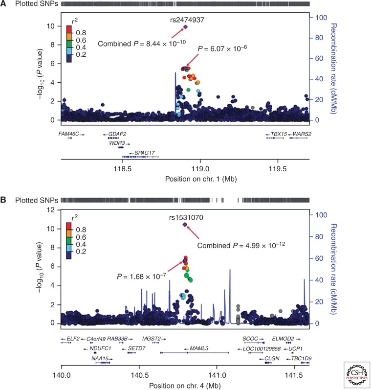
    1. Cordell HJ, Bentham J, Topf A, Zelenika D, Heath S, Mamasoula C, Cosgrove C, Blue G, Granados-Riveron J, Setchfield K, et al. 2013a. Genome-wide association study of multiple congenital heart disease phenotypes identifies a susceptibility locus for atrial septal defect at chromosome 4p16. Nat Genet 45: 822–824 - PMC - PubMed
    1. Cordell HJ, Topf A, Mamasoula C, Postma AV, Bentham J, Zelenika D, Heath S, Blue G, Cosgrove C, Granados Riveron J, et al. 2013b. Genome-wide association study identifies loci on 12q24 and 13q32 associated with tetralogy of Fallot. Hum Mol Genet 22: 1473–1481 - PMC - PubMed
    1. Cripe L, Andelfinger G, Martin LJ, Shooner K, Benson DW 2004. Bicuspid aortic valve is heritable. J Am Coll Cardiol 44: 138–143 - PubMed
    1. Dunham I, Kundaje A, Aldred SF, Collins PJ, Davis CA, Doyle F, Epstein CB, Frietze S, Harrow J, Kaul R, et al. 2012. An integrated encyclopedia of DNA elements in the human genome. Nature 489: 57–74 - PMC - PubMed

Publication types

MeSH terms

LinkOut - more resources