

Review
doi: 10.1152/physiol.00026.2013.
How could SNARE proteins open a fusion pore?
Affiliations
- PMID: 24985331
- PMCID: PMC4103061
- DOI: 10.1152/physiol.00026.2013
Item in Clipboard
Review
How could SNARE proteins open a fusion pore?
Physiology (Bethesda).
2014 Jul.
Abstract
The SNARE (Soluble NSF Attachment protein REceptor) complex, which in mammalian neurosecretory cells is composed of the proteins synaptobrevin 2 (also called VAMP2), syntaxin, and SNAP-25, plays a key role in vesicle fusion. In this review, we discuss the hypothesis that, in neurosecretory cells, fusion pore formation is directly accomplished by a conformational change in the SNARE complex via movement of the transmembrane domains.
©2014 Int. Union Physiol. Sci./Am. Physiol. Soc.
Conflict of interest statement
No conflicts of interest, financial or otherwise, are declared by the author(s).
Figures
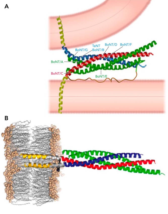
Structure of the SNARE complex A: hypothetical model for the structure of the trans SNARE complex as suggested by Ref. . The TM domains (yellow) and the SNAP-25 linker (brown) were not part of the crystal structure. B: structure of the post fusion cis SNARE complex, including the TM domains of syb2 and stx, shows helical extension of the SNARE domains into the membrane (56). Figure adapted from Refs. and and used with permission from Nature.
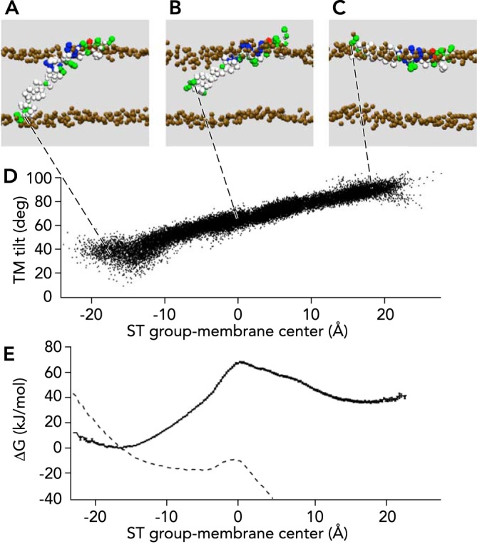
Energetics of syb2 tilting movement in the membrane Harmonic potentials were applied to S115/T116 (ST group) in state (A), generating directional movement through the hydrophobic core (B) to the extravesicular membrane-water interface (C). D: the change of the ST group position is accompanied by a corresponding change of the TM domain tilt angle. E: the free energy profile shows a maximum when the COOH terminus is located near the membrane center (black dots with error bars). The energy barrier is lowered to ∼3 kBT when an 80-pN force is applied in z direction (broken line). Image adapted from Ref. and used with permission.
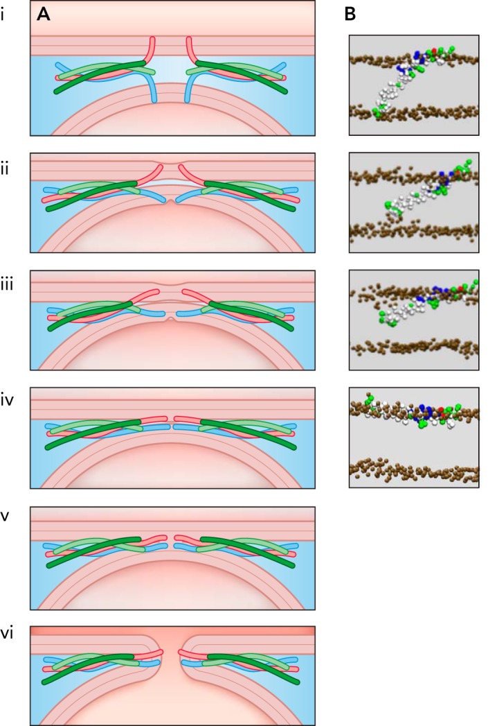
Model of TM domain movements Model of TM domain movements leading to fusion pore formation (A) and corresponding states of the syb2 TM domain from CG-MD simulations (35). The zippering of the SNARE complex will lead to helical extension, leading to force transfer and TM domain, tilt as indicated in states i–iv. Fusion pore formation (state vi) could proceed via the stalk state (state v) or could be induced via the transition from state iii to iv. Image adapted from Ref. and used with permission.
References
-
- Adams SR, Campbell RE, Gross LA, Martin BR, Walkup GK, Yao Y, Llopis J, Tsien RY. New biarsenical ligands and tetracysteine motifs for protein labeling in vitro and in vivo: synthesis and biological applications. J Am Chem Soc 124: 6063–6076, 2002 - PubMed
-
- An SJ, Almers W. Tracking SNARE complex formation in live endocrine cells. Science 306: 1042–1046, 2004 - PubMed
-
- Aoyagi K, Sugaya T, Umeda M, Yamamoto S, Terakawa S, Takahashi M. The activation of exocytotic sites by the formation of phosphatidylinositol 4,5-bisphosphate microdomains at syntaxin clusters. J Biol Chem 280: 17346–17352, 2005 - PubMed
-
- Brose N, Petrenko AG, Südhof TC, Jahn R. Synaptotagmin: a calcium sensor on the synaptic vesicle surface. Science 256: 1021–1025, 1992 - PubMed
Publication types
MeSH terms
Substances
Grants and funding
LinkOut - more resources
Full Text Sources
Other Literature Sources