Review
. 2014 Jul;29(4):286-98.
doi: 10.1152/physiol.00001.2014.

Germ cell transport across the seminiferous epithelium during spermatogenesis

Affiliations
Review

Germ cell transport across the seminiferous epithelium during spermatogenesis

Xiang Xiao et al. Physiology (Bethesda). 2014 Jul.

Abstract

Transport of germ cells across the seminiferous epithelium is crucial to spermatogenesis. Its disruption causes infertility. Signaling molecules, such as focal adhesion kinase, c-Yes, c-Src, and intercellular adhesion molecules 1 and 2, are involved in these events by regulating actin-based cytoskeleton via their action on actin-regulating proteins, endocytic vesicle-mediated protein trafficking, and adhesion protein complexes. We critically evaluate these findings and provide a hypothetical framework that regulates these events.


Conflict of interest statement

No conflicts of interest, financial or otherwise, are declared by the author(s).

Figures
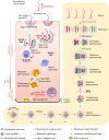
FIGURE 1.
FIGURE 1.
The biology of spermatogenesis Left: a schematic drawing that illustrates the cross section of the seminiferous tubule, the functional unit in the testis that produces ∼300–400 million spermatozoa per day in a man after puberty at ∼12 yr of age, which persists throughout his entire life. Spermatogenesis takes place in the seminiferous epithelium, which is composed of only Sertoli and germ cells, located above the tunica propria. Spermatogonia that reside near the basement membrane derived from spermatogonial stem cells (SSC) enter spermatogenesis and differentiate into As (Asingle), to be followed by A1–A4 and intermediate (In) spermatogonia, until type B spermatogonia transform to preleptotene spermatocytes, which are the germ cells that must be transported across the blood-testis barrier (BTB) at stage VIII of the epithelial cycle. The BTB also divides the seminiferous epithelium into the adluminal and the basal compartment. Meiosis I and II, and all the cellular events of postmeiotic development known as spermiogenesis and spermiation, all take place in the adluminal compartment. During these processes, millions of spermatozoa are formed efficiently, and developing germ cells are also being transported progressively from the basal to the adluminal compartment, and finally to the edge of the seminiferous tubule lumen, so that mature spermatozoa can be released at spermiation to enter the seminiferous tubule lumen for their eventual maturation in the epididymis. ES, ectoplasmic specialization.
FIGURE 2.
FIGURE 2.
The seminiferous epithelial cycle of spermatogenesis A unique feature of spermatogenesis is the cyclic association of germ cells with the Sertoli cell (annotated by “red” arrowhead) in the seminiferous epithelium, such as in the rat testis as illustrated in A, in which stages I–XIV can be defined (note: only I–XII and I–VI stages are found in the mouse and human testis, respectively). Each stage of the cycle shown in A illustrates unique association of specific germ cells and the Sertoli cell in the seminiferous epithelium, and the different germ cell types that are found in each stage are shown in B. For instance, in stage VIII of the epithelial cycle, which lasts for ∼29.1 h in the rat, step 19 spermatids line up near the tubule lumen to prepare for spermiation. It is also noted that preleptotene spermatocytes that derive from type B spermatogonia that first appear in stage VII are being transported across the BTB in stages VII–VIII. Thus, in stage VIII, only type A1 spermatogonia, preleptotene spermatocytes, pachytene spermatocytes, and steps 8 and 19 spermatids are found. Also, meiosis only takes place in stage XIV in the rat testis. In short, the entire epithelial cycle from I to XIV takes ∼12.9 days to complete. However, it takes a type A1 spermatogonium in stage II to become a step 19 spermatid in stage VIII through the epithelial cycle ∼4.5 times, which lasts for ∼58 days in the rat. A1, type A1 spermatogonium; A1m, type A1 spermatogonium undergoes mitosis; In, intermediate spermatogonium; Inm, intermediate spermatogonium undergoes mitosis; B, type B spermatogonium; Bm, type B spermatogonium undergoes mitosis; Pl, preleptotene spermatocyte; L, leptotene spermatocyte; Z, zygotene spermatocyte; P, pachytene spermatocyte; 1–19, step 1 to step 19 spermatids that are formed during spermiogenesis; ES, ectoplasmic specialization.
FIGURE 3.
FIGURE 3.
A schematic drawing that illustrates the concept of junction restructuring in the seminiferous epithelium in relation to spermatid transport during spermiogenesis and spermiation For the transport of spermatids across the seminiferous epithelium during the epithelial cycle of spermatogenesis, apical ectoplasmic specialization (ES) is the anchoring junction responsible for this cellular event. Left: this represents the state of an intact apical ES at the Sertoli-spermatid interface, such as in a stage VII tubule. Apical ES first appears in step 8 spermatids; once it forms, it replaces desmosome and gap junction as the only anchoring device to confer spermatid adhesion and polarity, and it persists until step 19 spermatids in stage VII to early stage VIII until spermiation takes place in late stage VIII of the epithelial cycle. Spermatids anchor to the Sertoli cell at the apical ES using integral membrane proteins, such as ICAM-2, nectins (nectin 3 is spermatid-specific, but nectin 2 is found in both Sertoli cells and spermatids), and most notably α6β1-integrin/laminin-333 (integrin is Sertoli cell-specific, whereas laminin is spermatid-specific). Apical ES is maintained via a network of actin filament bundles that are sandwiched between cisternae of endoplasmic reticulum and the Sertoli cell plasma membrane. The actin filament bundles, in turn, are maintained by actin bundling proteins, such as Eps8 and palladin, and also signaling molecules p-FAK-Tyr407, p-FAK-Tyr397, c-Yes, and c-Src. In early stage VIII, the expression of actin bundling proteins (e.g., Eps8 and palladin) is downregulated, and the expression of actin branching protein, the Arp2/3 complex, is induced. This combined effect changes the F-actin from its “bundled” to “branched/un-bundled” configuration efficiently. Such re-organization of F-actin network thus destabilizes the adhesion protein complexes, and the nonreceptor protein tyrosine kinases (e.g., c-Yes and c-Src) also facilitate endocytic vesicle-mediated protein trafficking, such as endocytosis, transcytosis, and/or recycling. The recycling and/or endosome-mediated degradation of integral membrane proteins at the apical ES further destabilizes spermatid adhesion, facilitating the release of sperm at spermiation. Furthermore, the transcytosed and recycled apical ES proteins facilitate the assembly of “new” apical ES when step 8 spermatids appear in stage VIII tubules (see FIGURE 2). In short, this effectively re-organizes actin filament bundles at the apical ES via the intricate spatiotemporal expression of actin bundling proteins (e.g., Eps8, palladin) and actin branching/un-bundling proteins (e.g., the Arp2/3 complex) mediated by p-FAK-Tyr407 and p-FAK-Tyr397, thus rapidly converting actin filaments from “bundled” and “branched/un-bundled” configuration and vice versa. These changes coupled with endocytic vesicle-mediated trafficking events mediated by c-Yes and c-Src provide the means to facilitate spermatid transport across the seminiferous epithelium during spermatogenesis.
FIGURE 4.
FIGURE 4.
A schematic drawing that illustrates the concept of junction restructuring at the blood-testis barrier (BTB) in relation to preleptotene spermatocyte transport across the immunological barrier during spermatogenesis Left: a preleptotene spermatocyte at stage VII of the epithelial cycle just arising from type B spermatogonium is located behind the intact BTB in a stage VII tubule. The BTB integrity is conferred by two arrays of actin filament bundles, which are also maintained by actin bundling proteins, such as Eps8 and palladin. BTB integrity is also conferred by adhesion protein complexes, such as occludin-ZO-1, N-cadherin-β-catenin, ICAM-1-ZO-1, as well as signaling proteins p-FAK-Tyr407, c-Yes, and c-Src. Similar to the apical ES, actin filament bundles at the basal ES are located between the cisternae of endoplasmic reticulum and the Sertoli cell plasma membrane. Middle: as described in the text, laminin fragments that are generated at the apical ES during spermiation via the action of MMP-2 on laminin chains possess biological activity to induce BTB restructuring, which is mediated by changes in the spatiotemporal expression of actin bundling proteins (e.g., Eps8, palladin), branched actin polymerization protein (e.g., the Arp2/3 complex which is activated by N-WASP to induce barbed end actin polymerization), and signaling proteins p-FAK-Tyr407, c-Yes, and c-Src. Also, soluble ICAM-1 (sICAM-1) is generated from ICAM-1 via the action of MMP-9, which promotes BTB restructuring. The net result of these changes induces reorganization of the F-actin network, causing actin filament bundles altered from a “bundled” to a “branched/unbundled” configuration, thereby destabilizing adhesive function of the TJ, basal ES, and gap junction, and also facilitating endocytic vesicle-mediated protein trafficking. Thus the “old” BTB above the preleptotene spermatocyte is disrupted. Right: at the same time the actin filament bundles are altered from a “bundled” to a “branched/unbundled” configuration, transcytosis and recycling have facilitated the assembly of a “new” BTB located at the basal region of the preleptotene spermatocyte. The preleptotene spermatocyte will be transformed into leptotene spermatocyte at stages IX–XI to be followed by zygotene spermatocyte at stages XII–XIII (FIGURE 2). Using such a mechanism, the immunological barrier can be maintained during the transport of preleptotene spermatocytes across the BTB at stage VIII of the epithelial cycle. ES, ectoplasmic specialization.

Similar articles

Cited by

References

    1. Amann RP. The cycle of the seminiferous epithelium in humans: a need to revisit? J Androl 29: 469–487, 2008 - PubMed
    1. Amann RP, Howards SS. Daily spermatozoal production and epididymal spermatozoal reserves of the human male. J Urol 124: 211–215, 1980 - PubMed
    1. Amann RP, Schanbacher BD. Physiology of male reproduction. J Anim Sci 57, Suppl 2: 380–403, 1983 - PubMed
    1. Arold ST. How focal adhesion kinase achieves regulation by linking ligand binding, localization and action. Curr Opin Struct Biol 21: 808–813, 2011 - PMC - PubMed
    1. Auharek SA, Avelar GF, Lara NLM, Sharpe RM, Franca LR. Sertoli cell numbers and spermatogenic efficency are increased in inducible nitric oxide synthase (iNOS) mutant-mice. Int J Androl 34: e621–e629, 2011 - PubMed

Publication types

LinkOut - more resources