Review
. 2014 Aug;24(8):472-8.
doi: 10.1016/j.tcb.2014.06.001. Epub 2014 Jul 4.

Hypoxia, lipids, and cancer: surviving the harsh tumor microenvironment

Affiliations
Review

Hypoxia, lipids, and cancer: surviving the harsh tumor microenvironment

Daniel Ackerman et al. Trends Cell Biol. 2014 Aug.

Abstract

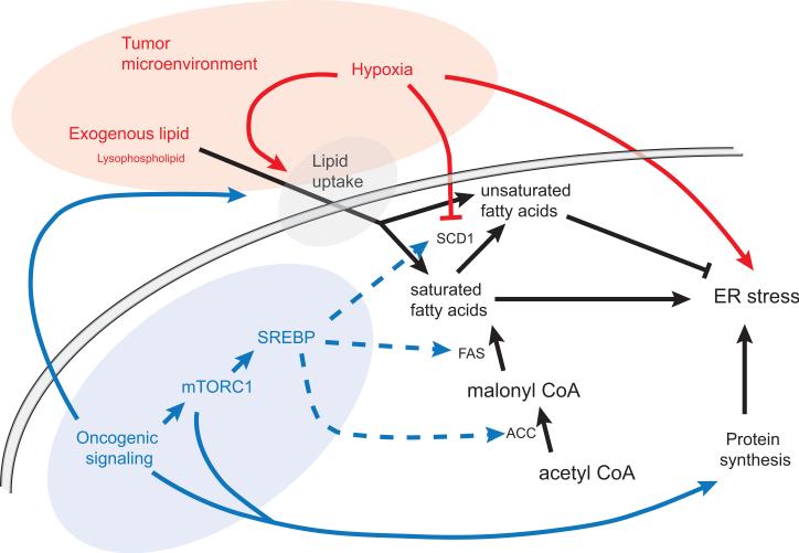
Solid tumors typically develop hostile microenvironments characterized by irregular vascularization and poor oxygen (O2) and nutrient supply. Whereas normal cells modulate anabolic and catabolic pathways in response to changes in nutrient availability, cancer cells exhibit unregulated growth even under nutrient scarcity. Recent studies have demonstrated that constitutive activation of growth-promoting pathways results in dependence on unsaturated fatty acids for survival under O2 deprivation. In cancer cells, this dependence represents a critical metabolic vulnerability that could be exploited therapeutically. Here we review how this dependence on unsaturated lipids is affected by the microenvironmental conditions faced by cancer cells.

Keywords: ER stress; SCD1; hypoxia; metabolism; unsaturated lipids.


Figures

Figure 1
Figure 1
Unsaturated lipids can be acquired by desaturation of de novo synthesized fatty acids or by uptake of exogenous lipid. Saturated fatty acids have been shown to promote ER stress while unsaturated fatty acids counteract the effect of saturated lipid. The tumor microenvironment can contribute to ER stress by inhibiting optimal protein folding and SCD1 desaturase activity. Hypoxia can also increase lipid uptake, which counteracts the effects of SCD1 inhibition. Oncogenic signaling increases fatty acid synthesis and protein synthesis. Elevated rates of protein synthesis have been shown to increase ER stress. Oncogenic changes, such as activating Ras mutations, also promote lipid uptake. SCD1: stearoyl-Coenzyme A desaturase 1 FAS: fatty acid synthase ACC: acetyl-CoA carboxylase
Figure 2
Figure 2
Increases in phospholipid saturation have been shown to activate the IRE1α and PERK branches of the UPR. The mechanisms of activation by lipid saturation are distinct from those triggered by unfolded protein accumulation. Conventional ER stress triggers IRE1α and PERK through either direct binding of unfolded proteins to the luminal domains of the sensors or through their interaction with the BiP chaperone protein [9]. While UPR activation by increased unfolded protein load occurs via the luminal domains of IREα and PERK, these domains are not required for activation by elevated lipid saturation. In addition, IRE1α activation by saturated lipids does not lead to the characteristic formation of IRE1α “foci” indicating that lipid-induced IRE1α activation does not lead to oligomerization.

References

    1. Hollien J, Weissman JS. Decay of endoplasmic reticulum-localized mRNAs during the unfolded protein response. Science. 2006;313:104–107. - PubMed
    1. Yoshida H, et al. XBP1 mRNA is induced by ATF6 and spliced by IRE1 in response to ER stress to produce a highly active transcription factor. Cell. 2001;107:881–891. - PubMed
    1. Upton JP, et al. IRE1alpha cleaves select microRNAs during ER stress to derepress translation of proapoptotic Caspase-2. Science. 2012;338:818–822. - PMC - PubMed
    1. Lerner AG, et al. IRE1alpha induces thioredoxin-interacting protein to activate the NLRP3 inflammasome and promote programmed cell death under irremediable ER stress. Cell metabolism. 2012;16:250–264. - PMC - PubMed
    1. Balkwill FR, et al. The tumor microenvironment at a glance. Journal of cell science. 2012;125:5591–5596. - PubMed