

Extra-nodal subcutaneous Hodgkin's-like lymphoma and subsequent regression in a cat
- PMID: 24985969
- PMCID: PMC10816795
- DOI: 10.1177/1098612X14541262
Extra-nodal subcutaneous Hodgkin's-like lymphoma and subsequent regression in a cat
Abstract
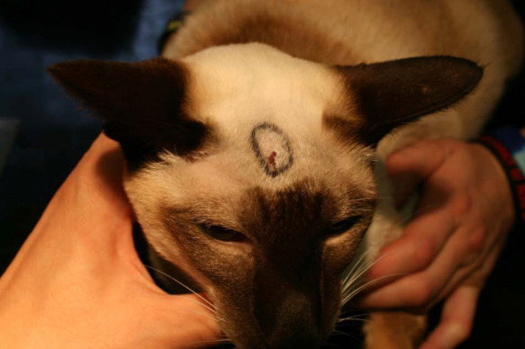
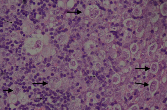
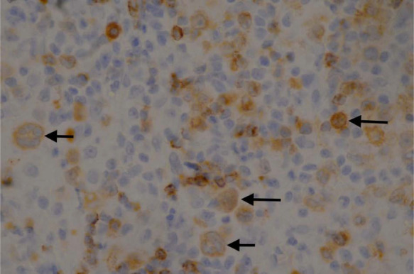
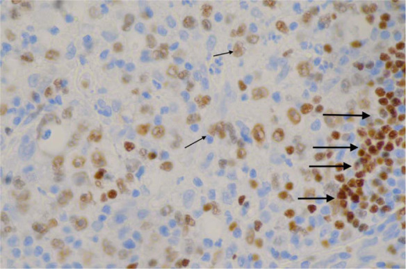
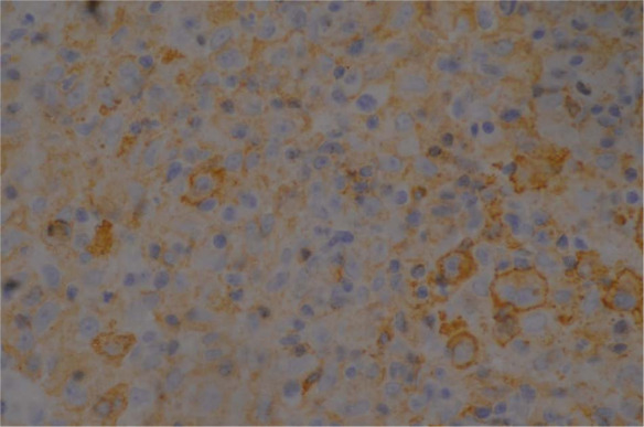
Hodgkin's-like lymphoma is a slow growing neoplasm, usually affecting the lymph nodes of the head and neck, which has been sporadically described in veterinary patients. This report describes the clinical and histopathological features in a 9-year-old male neutered Siamese cat that presented with a 6 week history of mid-dorsocranial swelling. Immunohistochemistry demonstrated positive staining for CD79a, paired box protein and B lymphocyte antigen-36, with variable, weak-to-moderate cytoplasmic staining for human leukocyte antigen-DR and CD18, and negative staining for antimacrophage antibody. The diagnosis based on incisional biopsy was Hodgkin's-like lymphoma; however, no evidence of neoplasia was found following wide surgical excision. This case report demonstrates two unreported items of note: the novel extranodal site of Hodgkin's-like lymphoma in a cat and tumour regression following initial biopsy. It is hypothesised that the surgical trauma of biopsying the lesion or the introduction of foreign material may have caused the regression of the neoplastic cells through induction of an anti-tumour immune or inflammatory response.
© ISFM and AAFP 2014.
Conflict of interest statement
The authors do not have any potential conflicts of interest to declare.
Figures

References
-
- Day MJ, Silkstona MA, Luck VM, et al. . T-cell-rich B-cell lymphoma in the cat. J Comp Pathol 1999; 120: 155–167. - PubMed
-
- Chittal SM, Brousset P, Voigt JJ, et al. . Large B-cell lymphoma rich in T-cells and simulating Hodgkin’s disease. Histopathology 1991; 19: 211–220. - PubMed
-
- Rodriguez J, Pug WC, Cabanillas F. T-cell-rich B-cell lymphoma. Blood 1993; 82: 1586–1589. - PubMed
-
- Walton RM, Hendrick MJ. Feline Hodgkin’s-like Lymphoma: 20 Cases (1992–1999). Vet Pathol 2001; 38: 504–511. - PubMed
-
- Jose BO, Koerner P, Spanos WJ, Jr, et al. . Hodgkin’s lymphoma in adults – clinical features. J Ky Med Assoc 2005; 103: 15–17. - PubMed
Publication types
MeSH terms
LinkOut - more resources
Full Text Sources
Other Literature Sources
Medical
Miscellaneous
