. 2014 Sep;5(9):692-703.
doi: 10.1007/s13238-014-0087-3. Epub 2014 Jul 2.

Molecular mechanism of SCARB2-mediated attachment and uncoating of EV71

Affiliations

Molecular mechanism of SCARB2-mediated attachment and uncoating of EV71

Minghao Dang et al. Protein Cell. 2014 Sep.

Abstract

Unlike the well-established picture for the entry of enveloped viruses, the mechanism of cellular entry of non-enveloped eukaryotic viruses remains largely mysterious. Picornaviruses are representative models for such viruses, and initiate this entry process by their functional receptors. Here we present the structural and functional studies of SCARB2, a functional receptor of the important human enterovirus 71 (EV71). SCARB2 is responsible for attachment as well as uncoating of EV71. Differences in the structures of SCARB2 under neutral and acidic conditions reveal that SCARB2 undergoes a pivotal pH-dependent conformational change which opens a lipid-transfer tunnel to mediate the expulsion of a hydrophobic pocket factor from the virion, a pre-requisite for uncoating. We have also identified the key residues essential for attachment to SCARB2, identifying the canyon region of EV71 as mediating the receptor interaction. Together these results provide a clear understanding of cellular attachment and initiation of uncoating for enteroviruses.


Figures

Figure 1
Figure 1
Overview of the structures and characteristics of SCARB2. (A) Schematic diagram of domain organization of SCARB2. TM, transmembrane domain; LD, luminal domain; CD, cytoplasmic domain. The overall structures of nSCARB2 (B) and aSCARB2 (C) are shown as cartoons. Domain I, II and III are colored in orange, violet and lemon, respectively. Glycans, cysteine residues and disulfide bonds are depicted as colored sticks. Helices α5 of nSCARB2 and aSCARB2 are highlighted in red and slate respectively. (D) Superposition of domain III of nSCARB2 and aSCARB2. The residues making ionic interactions and hydrophobic interactions are shown as colored sticks. The luminal tunnels of nSCARB2 (E) and aSCARB2 (F) were generated by HOLLOW (Ho and Gruswitz, 2008) software and are shown as blue mesh. See also Fig. S1 and Table S1
Figure 2
Figure 2
SCARB2-mediated expulsion of lipid from EV71 virions triggers conformational change of EV71 mature virions under acidic conditions. Competitive displacement of natural lipid from EV71 capsid proteins using [3H]-labeled sphingosine. (A) Time dependence of competitive [3H]-labeled sphingosine binding to EV71 mature virions. Purified EV71 mature virions were incubated with [3H]-labeled sphingosine (100 nmol/L) for the indicated times in PBS buffer (pH 7.4). EV71 virions were pelleted, washed for 3 times using PBS buffer (pH 7.4) and the radioactivity associated with them was estimated. Results shown are the mean ± SEM of three independent experiments. (B) Time dependence of [3H]-labeled sphingosine release from EV71 mature virions in the presence of SCARB2 under acidic conditions. EV71 mature virions bearing [3H]-labeled sphingosine were incubated with Ni-NTA beads in the absence or presence of an excess of recombinant SCARB2 (with a His tag) in acidic pH buffer (pH 5.0) at 37°C for the indicated times. Differential ultracentrifugation was used to pellet EV71 particles. Radioactivity and protein content was estimated. Results shown are the mean ± SEM of three independent experiments. The sedimentation coefficients of EV71 virions were measured by using analytical ultracentrifugation (AUC). (C) EV71 virions were suspended in neutral pH buffer (pH 7.4) or in acidic pH buffer (pH 5.0); (D) EV71 virions were incubated with SCARB2 under neutral condition (pH 7.4) or under acidic condition (pH 5.0) at 37°C for 1 h; (E) EV71 virions were pretreated using NLD compound, and then incubated with SCARB2 under neutral condition (pH 7.4) or under acidic condition (pH 5.0) at 37°C for 1 h. Assays carried out under neutral pH buffer (pH 7.4) and acidic condition (pH 5.0) are presented in blue and red curves, respectively. See also Fig. S2
Figure 3
Figure 3
Identification of binding interface between EV71 and SCARB2. (A) GST pull-down assay for detecting interactions of SCARB2 with peptides located on the outer surface of the EV71 particle in vitro. Glutathione-Sepharose beads mixed with approximately 5 μg of GST-peptide were incubated with SCARB2. After the beads were washed, proteins that bound to the beads were analyzed by 15% SDS-PAGE, followed by Western blot analysis. The positions of peptide-GST and SCARB2 are marked on the right. (B) Titration of EV71 mature virions (7 μmol/L) with synthesized peptide of SCARB2 (aa 146–166, 400 μmol/L). Raw injection heats are shown in the top panel and the corresponding specific binding isotherm (calculated from the integrated injection heats and normalized to moles of injectant) are shown in the bottom panels. The derived dissociation constant (Kd), stoichiometry parameter (N), and change in molar enthalpy (ΔH) and entropy (ΔS) are also shown. (C) Peptides around the “canyon” region of EV71 interact with SCARB2. Surface rendering of one icosahedral asymmetric unit (PDB code: 3VBH) of EV71. EV71 capsid protein VP1, VP2 and VP3 are colored in light blue, pale green and salmon, respectively. Peptides showing stronger interaction with SCARB2 are colored in red and peptides having a weaker affinity for SCARB2 in yellow. A 5-fold axis is shown as a black line and the pocket factor (cyan) indicated by a black arrow. See also Fig. S3 and Table S2
Figure 4
Figure 4
Roles of glycosylation of SCARB2 in EV71 binding and infection. (A) Pull-down assay for the interaction of SCARB2 or deglycosylated (DG) SCARB2 with EV71 mature virions in vitro. Ni-NTA beads mixed with approximately 3 μg of SCARB2 or deglycosylated SCARB2 were incubated with EV71 mature virions. Similar steps as Fig. 3A were carried out and the positions of SCARB2, DG-SCARB2 and EV71 are marked on the right. (B) Man8GlcNA2 at N325 extends to binding domain. The structure of Man8GlcNA2 and domain III are shown in the same format as in Fig. 1. (C) Electron density maps of Man8GlcNA2 at N325 of nSCARB2 (2FOFC map contoured at 1.0 σ). EV71-GFP was used to infect 293A-hSCARB2 cell line pretreated with or without PNGase F. Fluorescence of GFP was determined 16 h post infection and EV71 infectivity was calculated and normalized to the infectivity to 293A-hSCARB2 cell line without any treatments, which was considered as 100%. (D) The levels of SCARB2 expression on the cell membrane from 293A-hSCARB2 cell line pretreated with or without PNGase F were monitored by flow cytometry assay. (E) Binding affinity comparisons of EV71 to 293A-hSCARB2 cell line pretreated with or without PNGase F. (F) Infection efficiency of EV71 to 293A-hSCARB2 cell line pretreated with or without PNGase F. Results shown are the mean ± SEM of three independent experiments for panel (D–F). See also Fig. S4
Figure 5
Figure 5
Putative molecular mechanism of SCARB2-assisted attachment and uncoating of EV71. The models of complexes of nSCARB2 (A), aSCARB2 (C) and one icosahedral asymmetric unit of EV71 (PDB code: 3VBH). VP1, VP2, VP3 and SCARB2 are drawn in blue, green, red and violet respectively. Potential residues involved in the binding of EV71 with SCARB2 are shown as spheres. The luminal tunnel of SCARB2 and hydrophobic pocket in VP1 from EV71 are represented as blue meshes. Pocket factor is shown in sticks. (B) and (D) are an enlarged representation of the EV71-SCARB2 interface. See also Figs. S5 and S6
Figure 6
Figure 6
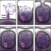
Cartoon of SCARB2-mediated EV71 entry. SCARB2 is inserted in the membrane with the ectodomain oriented towards the exofacial leaflet of the membrane. Pocket factor in VP1 is drawn as a “worm”. EV71 mature virion is shown as a purple icosahedron, and the deep-purple bigger icosahedron represents the EV71 uncoating intermediate. (A) EV71 in the process of attachment to the host cell membrane. (B) EV71 recognizes and interacts with cellular receptor SCARB2. (C) SCARB2 changes its conformation to open its “lipid-binding” tunnel at low pH value (<5.5) (upon internalization and transfer to the late endosome). (D) Expulsion of pocket factor from the viral capsid occurs. (E) Pocket factor is delivered to the membrane through the tunnel of SCARB2, meanwhile, EV71 undergoes a series of conformational changes as part of uncoating. (F) EV71 might dissociate from SCARB2 and form a channel in the membrane to release its RNA using the VP1 N-terminus and VP4

Comment in

  • The cellular receptor for enterovirus 71.
    Liu Y, Rossmann MG. Liu Y, et al. Protein Cell. 2014 Sep;5(9):655-7. doi: 10.1007/s13238-014-0092-6. Protein Cell. 2014. PMID: 25103897 Free PMC article. No abstract available.

References

    1. Adams PD, Afonine PV, Bunkoczi G, Chen VB, Davis IW, Echols N, Headd JJ, Hung L-W, Kapral GJ, Grosse-Kunstleve RW. PHENIX: a comprehensive Python-based system for macromolecular structure solution. Acta Crystallogr D Biol Crystallogr. 2010;66:213–221. doi: 10.1107/S0907444909052925. - DOI - PMC - PubMed
    1. Arita M, Koike S, Aoki J, Horie H, Nomoto A. Interaction of poliovirus with its purified receptor and conformational alteration in the virion. J Virol. 1998;72:3578–3586. - PMC - PubMed
    1. Bergelson JM, Coyne CB (2013) Picornavirus entry. In: Viral entry into host cells. Springer, New York, pp 24–41
    1. Chen P, Song Z, Qi Y, Feng X, Xu N, Sun Y, Wu X, Yao X, Mao Q, Li X. Molecular determinants of enterovirus 71 viral entry cleft around Gln-172 on VP1 protein interacts with variable region on scavenge receptor B 2. J Biol Chem. 2012;287:6406–6420. doi: 10.1074/jbc.M111.301622. - DOI - PMC - PubMed
    1. Cohen CJ, Shieh JT, Pickles RJ, Okegawa T, Hsieh J-T, Bergelson JM. The coxsackievirus and adenovirus receptor is a transmembrane component of the tight junction. Proc Natl Acad Sci USA. 2001;98:15191–15196. doi: 10.1073/pnas.261452898. - DOI - PMC - PubMed

Publication types

MeSH terms