

The influence of amalgam fillings on the detection of approximal caries by cone beam CT: in vitro study
- PMID: 24986630
- PMCID: PMC4170842
- DOI: 10.1259/dmfr.20130342
The influence of amalgam fillings on the detection of approximal caries by cone beam CT: in vitro study
Abstract
Objectives: The aim of this CBCT investigation on the detection of caries was to assess the influence of artefacts produced by the presence of amalgam fillings located in the vicinity.
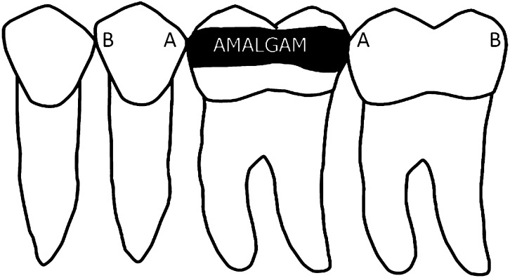
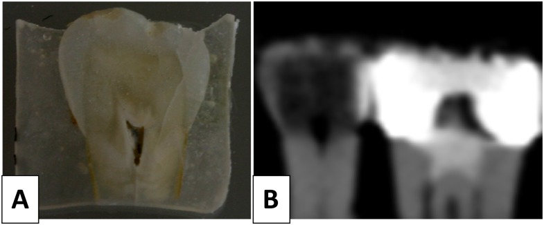
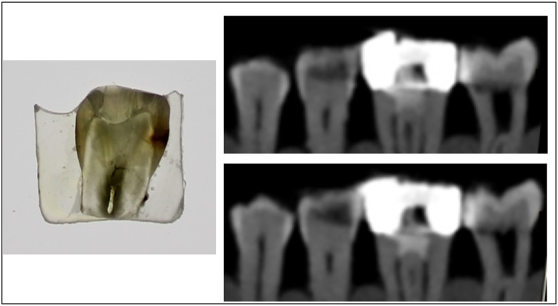
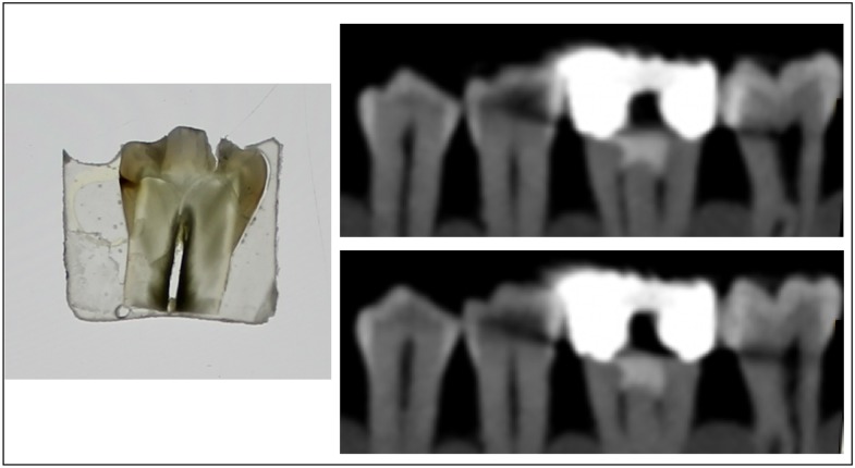
Methods: 102 non-cavitated pre-molar and molar teeth were placed in blocks of silicone with approximal contacts consisting of 3 sound or carious teeth and 1 mesial-occlusal-distal amalgam-filled tooth in-between. Radiographs of all the teeth were recorded using the CBCT system (NewTom™ 3G; QR Srl, Verona, Italy; field of view, 9 inches). Data from the CBCT unit were reconstructed and sectioned in the mesiodistal tooth plane. Images were evaluated twice by two observers, using a five-step confidence scale. After the CBCT examination, the teeth were individually sectioned in the mesiodistal direction with a diamond saw. Using a light microscope at ×40 magnification, the true morphological status of all approximal surfaces was established.
Results: Sensitivity of the CBCT for the detection of caries on surfaces located proximally and distally to an amalgam filing ranged from 0.27 to 0.30 for enamel and from 0.47 to 0.56 for dentin. Specificity values for enamel proximal and distal lesions were 0.48 and 0.53, respectively, for enamel and 0.33 to 0.38, respectively, for proximal and distal dentin cases. Intra-observer reliability was 0.84, and interobserver reliability was 0.49.
Conclusions: Owing to its low specificity, scans from a CBCT examination should not be used to determine the presence of demineralization of the tooth surface when amalgam fillings are present in the region of interest.
Keywords: CBCT; amalgam; artefacts; caries.
Figures
References
-
- Mozzo P, Procacci C, Tacconi A, Martini PT, Andreis IA. A new volumetric CT machine for dental imaging based on the cone-beam technique: preliminary results. Eur Radiol 1998; 8: 1558–64. - PubMed
-
- Kayipmaz S, Sezgin ÖS, Saricaoğlu ST, Çan G. An in vitro comparison of diagnostic abilities of conventional radiography, storage phosphor, and cone beam computed tomography to determine occlusal and approximal caries. Eur J Radiol 2011; 80: 478–82. - PubMed
LinkOut - more resources
Full Text Sources
Other Literature Sources
Miscellaneous
