

Regulation of a LATS-homolog by Ras GTPases is important for the control of cell division
- PMID: 24986648
- PMCID: PMC4120859
- DOI: 10.1186/1471-2121-15-25
Regulation of a LATS-homolog by Ras GTPases is important for the control of cell division
Abstract
Background: Nuclear Dbf-related/large tumor suppressor (NDR/LATS) kinases have been shown recently to control pathways that regulate mitotic exit, cytokinesis, cell growth, morphological changes and apoptosis. LATS kinases are core components of the Hippo signaling cascade and important tumor suppressors controlling cell proliferation and organ size in flies and mammals, and homologs are also present in yeast and Dictyostelium discoideum. Ras proto-oncogens regulate many biological functions, including differentiation, proliferation and apoptosis. Dysfunctions of LATS kinases or Ras GTPases have been implicated in the development of a variety of cancers in humans.
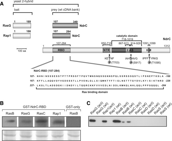
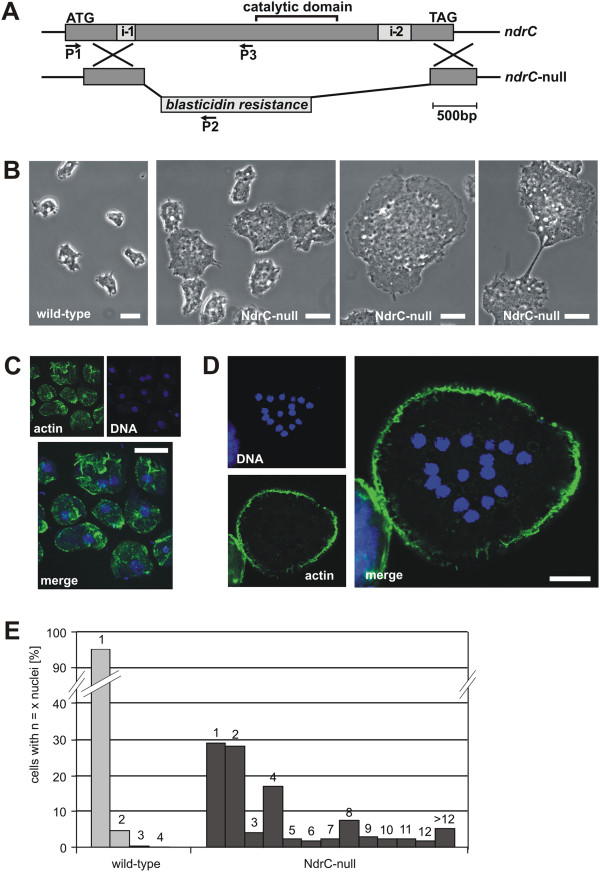
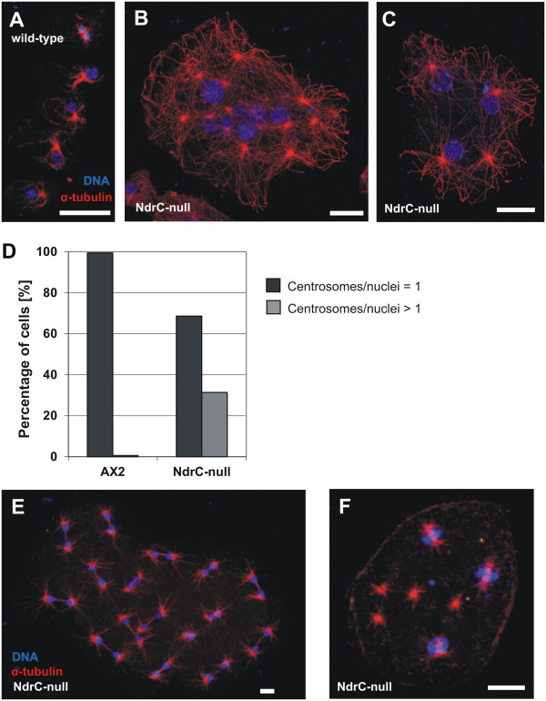
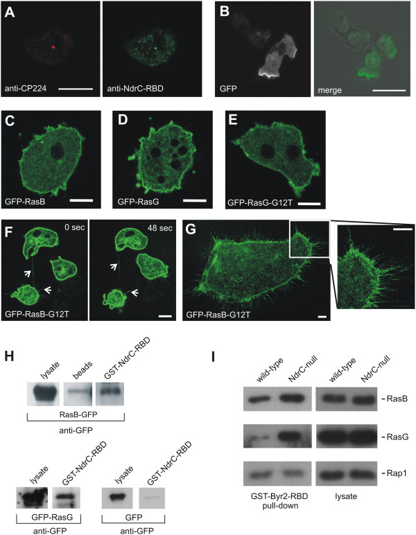
Results: In this study we used the model organism Dictyostelium discoideum to analyze the functions of NdrC, a homolog of the mammalian LATS2 protein, and present a novel regulatory mechanism for this kinase. Deletion of the ndrC gene caused impaired cell division and loss of centrosome integrity. A yeast two-hybrid analysis, using activated Ras proteins as bait, revealed NdrC as an interactor and identified its Ras-binding domain. Further in vitro pull-down assays showed that NdrC binds RasG and RasB, and to a lesser extent RasC and Rap1. In cells lacking NdrC, the levels of activated RasB and RasG are up-regulated, suggesting a functional connection between RasB, RasG, and NdrC.
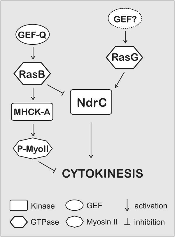
Conclusions: Dictyostelium discoideum NdrC is a LATS2-homologous kinase that is important for the regulation of cell division. NdrC contains a Ras-binding domain and interacts preferentially with RasB and RasG. Changed levels of both, RasB or RasG, have been shown previously to interfere with cell division. Since a defect in cell division is exhibited by NdrC-null cells, RasG-null cells, and cells overexpressing activated RasB, we propose a model for the regulation of cytokinesis by NdrC that involves the antagonistic control by RasB and RasG.
Figures
Similar articles
-
Functional overlap of the dictyostelium RasG, RasD and RasB proteins.J Cell Sci. 2000 Apr;113 ( Pt 8):1427-34. doi: 10.1242/jcs.113.8.1427. J Cell Sci. 2000. PMID: 10725225
-
Rap1 activation in response to cAMP occurs downstream of ras activation during Dictyostelium aggregation.J Biol Chem. 2008 Apr 18;283(16):10232-40. doi: 10.1074/jbc.M707459200. Epub 2008 Jan 7. J Biol Chem. 2008. PMID: 18180289
-
Degradation of activated K-Ras orthologue via K-Ras-specific lysine residues is required for cytokinesis.J Biol Chem. 2014 Feb 14;289(7):3950-9. doi: 10.1074/jbc.M113.531178. Epub 2013 Dec 13. J Biol Chem. 2014. PMID: 24338482 Free PMC article.
-
LATS tumor suppressor: a new governor of cellular homeostasis.Cell Cycle. 2010 Oct 1;9(19):3892-903. doi: 10.4161/cc.9.19.13386. Epub 2010 Oct 20. Cell Cycle. 2010. PMID: 20935475 Review.
-
Cytoskeletal regulation by Dictyostelium Ras subfamily proteins.J Muscle Res Cell Motil. 2002;23(7-8):729-36. doi: 10.1023/a:1024471527153. J Muscle Res Cell Motil. 2002. PMID: 12952071 Review.
Cited by
-
Quantitative imaging of Rac1 activity in Dictyostelium cells with a fluorescently labelled GTPase-binding domain from DPAKa kinase.Histochem Cell Biol. 2016 Sep;146(3):267-79. doi: 10.1007/s00418-016-1440-9. Epub 2016 Apr 28. Histochem Cell Biol. 2016. PMID: 27126594
-
The small GTPases Ras and Rap1 bind to and control TORC2 activity.Sci Rep. 2016 May 13;6:25823. doi: 10.1038/srep25823. Sci Rep. 2016. PMID: 27172998 Free PMC article.
-
The Hippo Tumor Suppressor Pathway (YAP/TAZ/TEAD/MST/LATS) and EGFR-RAS-RAF-MEK in cancer metastasis.Genes Dis. 2019 Dec 5;8(1):48-60. doi: 10.1016/j.gendis.2019.11.003. eCollection 2021 Jan. Genes Dis. 2019. PMID: 33569513 Free PMC article. Review.
-
The Dictyostelium Centrosome.Cells. 2021 Oct 5;10(10):2657. doi: 10.3390/cells10102657. Cells. 2021. PMID: 34685637 Free PMC article. Review.
-
CDK5RAP2 Is an Essential Scaffolding Protein of the Corona of the Dictyostelium Centrosome.Cells. 2018 Apr 23;7(4):32. doi: 10.3390/cells7040032. Cells. 2018. PMID: 29690637 Free PMC article.
References
-
- Hergovich A, Stegert MR, Schmitz D, Hemmings BA. NDR kinases regulate essential cell processes from yeast to humans. Nat Rev Mol Cell Biol. 2006;7(4):253–264. - PubMed
-
- Hergovich A, Cornils H, Hemmings BA. Mammalian NDR protein kinases: from regulation to a role in centrosome duplication. Biochim Biophys Acta. 2008;1784(1):3–15. - PubMed
-
- Hergovich A, Hemmings BA. Mammalian NDR/LATS protein kinases in hippo tumor suppressor signaling. Biofactors. 2009;35(4):338–345. - PubMed
Publication types
MeSH terms
Substances
LinkOut - more resources
Full Text Sources
Other Literature Sources
Molecular Biology Databases
