Review
. 2014 Jul;94(3):859-907.
doi: 10.1152/physrev.00037.2013.

Interstitial cells: regulators of smooth muscle function

Affiliations
Review

Interstitial cells: regulators of smooth muscle function

Kenton M Sanders et al. Physiol Rev. 2014 Jul.

Abstract

Smooth muscles are complex tissues containing a variety of cells in addition to muscle cells. Interstitial cells of mesenchymal origin interact with and form electrical connectivity with smooth muscle cells in many organs, and these cells provide important regulatory functions. For example, in the gastrointestinal tract, interstitial cells of Cajal (ICC) and PDGFRα(+) cells have been described, in detail, and represent distinct classes of cells with unique ultrastructure, molecular phenotypes, and functions. Smooth muscle cells are electrically coupled to ICC and PDGFRα(+) cells, forming an integrated unit called the SIP syncytium. SIP cells express a variety of receptors and ion channels, and conductance changes in any type of SIP cell affect the excitability and responses of the syncytium. SIP cells are known to provide pacemaker activity, propagation pathways for slow waves, transduction of inputs from motor neurons, and mechanosensitivity. Loss of interstitial cells has been associated with motor disorders of the gut. Interstitial cells are also found in a variety of other smooth muscles; however, in most cases, the physiological and pathophysiological roles for these cells have not been clearly defined. This review describes structural, functional, and molecular features of interstitial cells and discusses their contributions in determining the behaviors of smooth muscle tissues.


Figures

FIGURE 1.
FIGURE 1.
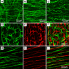
A–C: c-Kit+ ICC-MY in mouse, monkey, and human gastric antrums, respectively. Note similarities in the structural organization of ICC in all three species. D–F: c-Kit+ ICC-MY (D, green) and PDGFRα+-MY (fibroblast-like) cells (E, red) are distinct populations of cells. These panels show cells from murine colon. Merged images from D and E are shown in F. G–I: intramuscular ICC (ICC-IM; G, c-Kit is green) in the monkey gastric fundus. ICC-IM form close associations with enteric motor nerve processes (nNOS+ motor neuron processes are shown in H, red). I: merged image of G and H. Scale bar in F applies to D–F, and scale bar in I applies to G–I. (Authors are grateful to Dr. Masaaki Kurahashi for images in D–F and Dr. Peter Blair for images in B and G–I.)
FIGURE 2.
FIGURE 2.
Close connections between nerve varicosity and ICC (C1) and gap junction with smooth muscle cell in rat gastric antrum (183). Mitochondria, rough endoplasmic reticulum, and caveolae (arrowheads) are plentiful in ICC. Gap junction exists with adjacent smooth muscle cell (arrow). Very close contact is formed between nerve varicosity (N) and the ICC. Scale bar is 2 μm. Inset: higher magnification of the gap junction between ICC and smooth muscle cell. Scale bar in inset is 0.2 μm. [From Ishikawa et al. (183), with kind permission from Springer Science and Business Media.]
FIGURE 3.
FIGURE 3.
Structures of the SIP syncytium. A: electron micrograph (originally provided by Professor Terumasa Komuro) showing three types of cells in close proximity to nerve bundles (NB) and varicosities of enteric motor neurons (*): smooth muscle cells, ICC, and PDGFRα+ cells. These cells are electrically coupled (gap junctions not shown in this image), forming an electrical syncytium known as the SIP syncytium. In some cases, very close contacts (<20 nm; black arrow in inset) can be found between nerve varicosities and ICC or smooth muscle cells. Scale bars are 0.5 μm in main image and 0.2 μm in inset. B: depiction of SIP syncytium (with dotted line showing approximate cut of a thin section such as shown in A). Smooth muscle cells, ICC, and PDGFRα+ cells are arranged around projections of excitatory and inhibitory enteric motor neurons. [Redrawn from Sanders et al. (329). Copyright 2010 The Authors. Journal compilation Copyright 2010 The Physiological Society.]
FIGURE 4.
FIGURE 4.
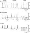
Electrical slow waves recorded from smooth muscle cells in various regions of the gastrointestinal (GI) tracts of 3 species. Slow waves consist of a relatively abrupt upstroke depolarization followed by a stable or slowly changing plateau phase. Although frequencies and amplitudes of slow waves vary in different tissues, note the regularity of duration and frequency in muscles from each region and species. The activity shown is integrated electrical behavior: a summation of slow waves generated by ICC, conducted to smooth muscle cells, and responses of smooth muscle cells. Depolarization of smooth muscle cells can activate voltage-dependent Ca2+ channels and generate Ca2+ action potentials. Ca2+ action potentials are elicited in muscles of the small intestine and colon during periods of enhanced excitation. Action potentials are not typical in gastric muscles, except in the terminal portion of the antrum and pylorus. Note differences in resting membrane potentials (most negative potentials between slow waves) in different regions. Regulation of membrane potential sets the excitability of these muscles and the response elicited by slow wave activity. Dotted lines were drawn at −40 mV, the approximate threshold for appreciable activation of L-type Ca2+ currents and excitation-contraction coupling. Note that slow waves are membrane potential fluctuations from negative potentials where open probability of voltage-dependent Ca2+ channels is very low to potentials where open probabilities increase and generate Ca2+ entry. Thus slow waves are an intrinsic mechanism organizing the motor output of GI muscles into phasic contractions. Propagation of slow waves in ICC networks generates segmental and peristaltic contractions. [From Sanders et al. (331).]
FIGURE 5.
FIGURE 5.
Loss of ICC-MY in small intestine of W/WV mice (404). A: networks of ICC-MY and ICC-DMP of the small intestine are shown in this whole-mount labeled with anti-c-Kit antibodies. B: loss of ICC-MY, but sparsely labeled ICC-DMP, running parallel to the circular muscle layer, remain in W/WV muscles. C: omnipresent slow wave activity recorded from wild-type muscles. D: loss of slow waves in small intestinal muscles of W/WV mice. Note also the significant depolarization that occurs in W/WV muscles. Recordings in C and D were from circular muscle cells.
FIGURE 6.
FIGURE 6.
Propagation of slow waves from ICC-MY to circular smooth muscle cells in the guinea pig stomach (trace was provided by Professor David Hirst). In the experiment illustrated, an ICC-MY was impaled (electrode 1) and a circular smooth muscle cell was impaled simultaneously. Traces above show slow wave events of large amplitude in ICC-MY and attenuated amplitude in smooth muscle cell. Note slight delay in activation of slow wave in smooth muscle cell.
FIGURE 7.
FIGURE 7.
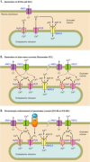
A model of electrical rhythmicity based on studies of freshly isolated ICC of the murine small intestine (433, 436, 437). Figures show portion of plasma membrane in close association with Ca2+ release mechanisms of the endoplasmic reticulum (ER). 1: The basic event of electrical rhythmicity is spontaneous transient inward currents (STICs) elicited by localized release of Ca2+ from stores. IP3-gated channels (IP3R) are the main source of Ca2+ driving activation of Ca2+-activated Cl channels (ANO1) in the plasma membrane to generate STICs, but ryanodine receptor (RyR) may also contribute. Ca2+ is recovered into stores via active Ca2+ pumping into the ER. STICs generate spontaneous transient depolarizations (STDs), the voltage response to the inward currents. 2: The second phase of rhythmicity is activation of T-type voltage-dependent Ca2+ channels (VDCC) in response to the STDs caused by STICs. Ca2+ entry synchronizes Ca2+ release in other regions of membrane, leading to synchronized opening of ANO1 channels throughout the cell. This generates slow wave currents. Depolarization also activates VDCC in adjacent electrically coupled cells, leading to cell-to-cell active propagation of slow waves in ICC networks. 3: In the 3rd phase of rhythmicity, binding of chronotropic agonists (such as ACh in this example) to G protein-coupled receptors (type 3 muscarinic receptor, M3, in this example) leads to IP3 formation and increased frequency and amplitude of STICs. More frequent STICs and greater amplitude depolarizations increase the likelihood of activation of whole cell slow wave currents. The steps in this mechanism are deduced from studies on ICC of the murine small intestine. Pharmacological data suggest that the generalized mechanism proposed applies to other GI muscles (e.g., dependence upon Ca2+-activated Cl currents to generate STICs and slow wave currents); however, the mechanism for regulation of frequency is poorly understood and appears to vary in ICC from different regions of the GI tract and the mechanism for voltage-dependent Ca2+ entry and/or release, necessary for slow wave propagation, appears to vary. For example, slow waves recorded from different regions of the murine GI tract and in other species display a range of sensitivities to extracellular Ca2+, Ni2+, and dihydropyridines. The ionic and molecular explanations for the diversity in the slow wave mechanism and for regulation of frequency are not fully understood.
FIGURE 8.
FIGURE 8.
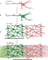
Active propagation of slow waves. 1: The initiating step occurs by localized Ca2+ transients (elevated Ca2+ depicted by green color in all cells; arrow), initiating STICs in an ICC. 2: Depolarization caused by STICs activates Ca2+ entry and Ca2+-induced Ca2+ release raising Ca2+ throughout the ICC and activating whole cell slow wave current. 3: Depolarization causes active propagation of slow waves through the ICC network (horizontal black arrow shows direction of propagation), and slow wave currents are activated cell to cell, as the wavefront spreads. 4: As slow waves propagate through the ICC network, they conduct passively into electrically coupled smooth muscle cells (SMCs). Slow waves depolarize SMCs and activate L-type Ca2+ channels. Ca2+ entry (green) triggers SMC contraction. Spread of slow waves in the ICC network leads spread of contractions necessary for segmental and peristaltic contractions.
FIGURE 9.
FIGURE 9.
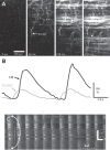
Spread of Ca2+ transients in the ICC network of murine small intestine. ICC were loaded with fluo 4. Sequence of video images shows development of Ca2+ transient in ICC-MY and spread of wave front from upper left quadrant to whole image. Streaks of fluorescence are longitudinal smooth muscle cells (LM) that are above the ICC-MY network. Note that ICC fire Ca2+ transients prior to events in LM. Ca2+ transients in regions of interest in ICC-MY and LM cells are shown as traces as a function of time in the bottom right of figure. Spatiotemporal maps were constructed from repetitive Ca2+ transients sweeping through the ICC-MY network. [Redrawn from Hennig et al. (147). Copyright 2010 Blackwell Publishing Ltd.]
FIGURE 10.
FIGURE 10.
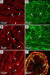
Interstitial cells in bladder and oviduct. A–C: PDGFRα+ cells in the detrusor muscle of cynomolgus monkey (Macaca fascicularis) bladder. A and B: cryostat cross sections through the detrusor wall. At least two populations of PDGFRα+ cells were observed: cells within (arrowheads) and surrounding detrusor muscle bundles (arrows). PDGFRα+ cells also appeared to bridge between adjacent muscle bundles (*). C: flat-profile cryostat section through the detrusor. PDGFRα+ cells located within (arrowheads) and between (arrows) muscle bundles are present. D–F: ICC and PDGFRα+ cells within the mouse and cynomolgus monkey oviduct. A and B: ICC within the myosalpinx of mouse (D) and monkey (E) form an anastomosing network of cells (arrows). These cells have been shown to provide electrical pacemaker function for propulsive contractions in the oviduct (see text for details). F: double labeling of PDGFRα+ cells in a cross section of the isthmus region of the mouse oviduct. Tissue comes from Pdgfratm11(EGFP)Sor/J mice that have constitutive expression of eGFP in nuclei driven off the Pdgfra promoter. The tissues were double-labeled with antibodies against PDGFRα. Thus the soma and cytoplasmic processes of PDGFRα+ cells are labeled with antibody (red), and nuclei of PDGFRα+ cells are green. A dense population of these cells were observed within the myosalpinx (arrowheads) and within the submucosa and endosalpinx (arrows) of the oviduct. The specific functions of these cells have not been determined. Scale bars are as indicated in each panel. (Authors are grateful to Dr. Rose Ellen Dixon for images shown in D and E.)

Comment in

References

    1. Abe H. The mammalian oviductal epithelium: regional variations in cytological and functional aspects of the oviductal secretory cells. Histol Histopathol 11: 743–768, 1996 - PubMed
    1. Abe Y, Tomita T. Cable properties of smooth muscle. J Physiol 196: 87–100, 1968 - PMC - PubMed
    1. Adams CE. Rate of sperm transport in the female reproductive tract of the rabbit. J Endocrinol 13: xxi–xxii, 1956 - PubMed
    1. Alberti E, Mikkelsen HB, Wang XY, Diaz M, Larsen JO, Huizinga JD, Jimenez M. Pacemaker activity and inhibitory neurotransmission in the colon of Ws/Ws mutant rats. Am J Physiol Gastrointest Liver Physiol 292: G1499–G1510, 2007 - PubMed
    1. Allix S, Reyes-Gomez E, Aubin-Houzelstein G, Noel D, Tiret L, Panthier JJ, Bernex F. Uterine contractions depend on KIT-positive interstitial cells in the mouse: genetic and pharmacological evidence. Biol Reprod 79: 510–517, 2008 - PubMed

Publication types