Clinical Trial
. 2014 Sep 1;20(17):4459-70.
doi: 10.1158/1078-0432.CCR-13-3361. Epub 2014 Jul 1.

p53MVA therapy in patients with refractory gastrointestinal malignancies elevates p53-specific CD8+ T-cell responses

Affiliations
Clinical Trial

p53MVA therapy in patients with refractory gastrointestinal malignancies elevates p53-specific CD8+ T-cell responses

Nicola R Hardwick et al. Clin Cancer Res. .

Erratum in

Abstract

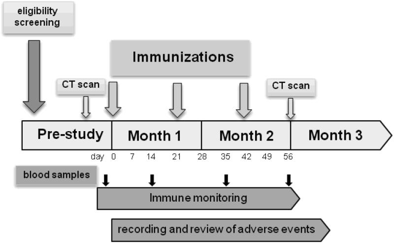
Purpose: To conduct a phase I trial of a modified vaccinia Ankara (MVA) vaccine delivering wild-type human p53 (p53MVA) in patients with refractory gastrointestinal cancers.

Experimental design: Three patients were vaccinated with 1.0×10(8) plaque-forming unit (pfu) p53MVA followed by nine patients at 5.6×10(8) pfu. Toxicity was classified using the NCI Common Toxicity Criteria and clinical responses were assessed by CT scan. Peripheral blood samples were collected pre- and post-immunization for immunophenotyping, monitoring of p53MVA-induced immune response, and examination of PD1 checkpoint inhibition in vitro.

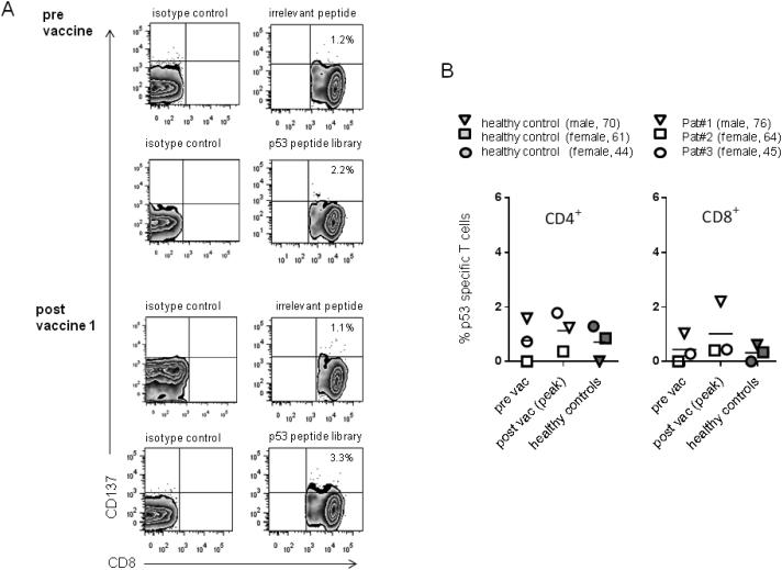
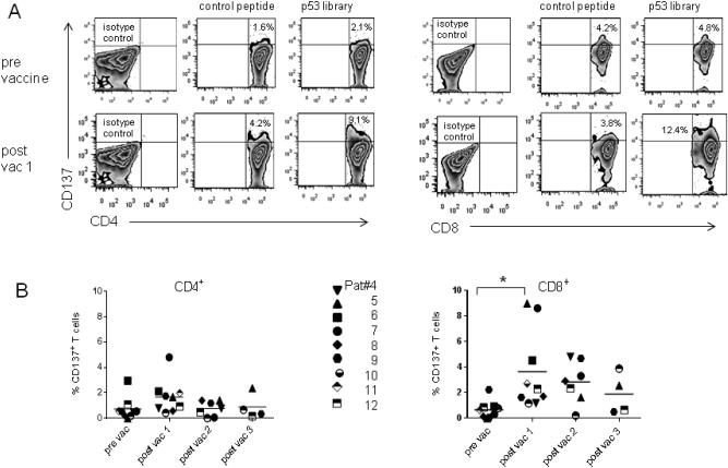
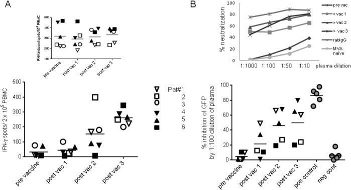
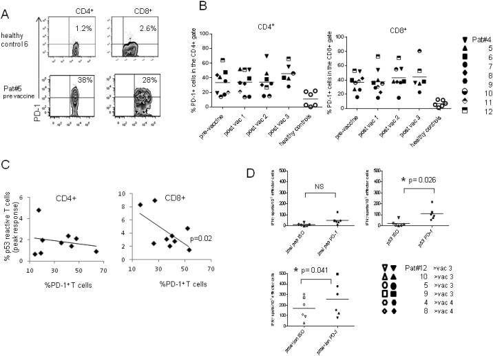
Results: p53MVA immunization was well tolerated at both doses, with no adverse events above grade 2. CD4+ and CD8+ T cells showing enhanced recognition of a p53 overlapping peptide library were detectable after the first immunization, particularly in the CD8+ T-cell compartment (P=0.03). However, in most patients, this did not expand further with the second and third immunization. The frequency of PD1+ T cells detectable in patients' peripheral blood mononuclear cells (PBMC) was significantly higher than in healthy controls. Furthermore, the frequency of PD1+ CD8+ T cells showed an inverse correlation with the peak CD8+ p53 response (P=0.02) and antibody blockade of PD1 in vitro increased the p53 immune responses detected after the second or third immunizations. Induction of strong T-cell and antibody responses to the MVA backbone were also apparent.

Conclusion: p53MVA was well tolerated and induced robust CD8+ T-cell responses. Combination of p53MVA with immune checkpoint inhibition could help sustain immune responses and lead to enhanced clinical benefit.


Figures

Figure 1
Figure 1
Clinical Trial Schema: vaccination and phlebotomy schedule for a p53MVA phase I clinical trial.
Figure 2
Figure 2. T cells collected from healthy donors and GI cancer patients receiving a subtherapeutic dose of p53MVA show low levels of p53 reactivity, despite in vitro stimulation
Patient T cells were co-cultured for 5 days with p53 expressing APC and 10U/ml IL-2. Stimulated cells were then washed and reseeded in fresh media for 2 days. After a final, overnight stimulation with a p53-peptide library, the percentage of CD4+ or CD8+ T cells expressing the activation marker CD137 was determined. Comparative analysis using PBMC from three unvaccinated, matched healthy controls were carried out in parallel. Panel A shows representative flow cytometry plots of the CD8+ population, pre and post vaccination 1 from Patient#1. Panel B shows the peak post-vaccine p53 response compared to pre-vaccine for both CD4+ and CD8+ T cells in patients 1-3. To control for non-specific stimulation % CD137+ T cells in response to p53 peptide - % CD137+ T cells in response to an irrelevant peptide were plotted. Each patient (open symbols) and the corresponding matched control (closed symbols) is represented by a different symbol.
Figure 3
Figure 3. Vaccination with high dose p53MVA transiently increases the frequency of p53-reactive T cells in the peripheral blood of GI cancer patients
Blood samples were collected pre-vaccination and two weeks after each p53MVA immunization. Purified CD3+ cells were expanded in vitro for 7 days prior to an overnight stimulation with a p53 peptide library or control peptides. The T cell activation marker, CD137, was used as a measure of T cell responsiveness and quantified by flow cytometry. Panel A shows representative flow cytometry plots from Patient#7. Panel B shows the % CD137+ T cells in response to p53 peptide - %CD137+ T cells in response to an irrelevant peptide in the CD4+ or CD8+ populations. Patients 4-12 are each represented by a different symbol. The increase in % of p53 responsive CD8+ T cells post-vaccine 1 compared to pre-vaccine values was statistically significant (p =0.03 ).
Figure 4
Figure 4. p53MVA immunization induces anti-MVA neutralizing antibody and T cell responses in advanced GI cancer patients
Panel A shows the T cell response from 3 low dose patients (open symbols) and 3 high dose patients (closed symbols) against VenusMVA. PBMC tested IFN-γ ELISPOT assays for reactivity to media alone, VenusMVA or PHA. Values plotted represent the mean number of spots from PBMC incubated with VenusMVA – the mean number of spots from PBMC seeded in media alone (background). The number of IFN-γ spots from PHA stimulated PBMC are shown separately above. Panel B shows the % neutralization of the Venus signal by plasma collected from the same patients. This was assessed by incubating the VenusMVA virus with patient or control plasma prior to infection of a permissive cell line. Venus expression was determined by flow cytometry. Data is shown for only 5 patients (Pat#3 plasma samples were unevaluable in this assay). Plasma from MVA naïve individuals and purified IgG from vaccinia vaccinated rabbits were included as negative and positive controls respectively. % neutralization obtained at 1:100 dilution are shown, with a representative dilution curve for Pat#1 shown above.
Figure 5
Figure 5. Advanced GI cancer patients show higher frequency of PD-1+ T cells than healthy controls both pre and post-immunization with high dose p53MVA
Peripheral blood T cells collected pre- and post-immunization were assessed by flow cytometry for the expression of the programmed cell death receptor PD-1. Panel A shows representative plots from a patient and healthy control. Quadrant gates were set according to isotype control staining. Panel B shows the % PD-1+ T cells in the CD4+ and CD8+ populations at different times in the immunization schedule. Different patients are represented by different symbols. Panel C shows an inverse relationship between the frequency of PD-1+ T cells pre-immunization and the peak p53-reactive T cells in the CD4+ and CD8+ populations. Correlation in the CD8+ population reached statistical significance (p=0.02). Figure 5D shows the reactivity of effector cells after in vitro expansion in the presence of αPD-1 (closed symbols) or isotype control (open symbols). Stimulated cells were tested in IFN-γ ELISPOT assays against control peptide, p53 library, VenusMVA and PMA + Ionomycin. Data shown is from lymphocytes obtained after 2 or 3 vaccinations.

References

    1. Katz SC, Donkor C, Glasgow K, Pillarisetty VG, Gonen M, Espat NJ, et al. T cell infiltrate and outcome following resection of intermediate-grade primary neuroendocrine tumours and liver metastases. HPB : the official journal of the International Hepato Pancreato Biliary Association. 2010;12:674–83. - PMC - PubMed
    1. Deschoolmeester V, Baay M, Van Marck E, Weyler J, Vermeulen P, Lardon F, et al. Tumor infiltrating lymphocytes: an intriguing player in the survival of colorectal cancer patients. BMC immunology. 2010;11:19. - PMC - PubMed
    1. Topalian SL, Hodi FS, Brahmer JR, Gettinger SN, Smith DC, McDermott DF, et al. Safety, activity, and immune correlates of anti-PD-1 antibody in cancer. N Engl J Med. 2012;366:2443–54. - PMC - PubMed
    1. Cheever MA, Higano C. PROVENGE (Sipuleucel-T) in Prostate Cancer: The First FDA Approved Therapeutic Cancer Vaccine. Clin Cancer Res. 2011;17:3520–6. - PubMed
    1. Hodi FS. Overcoming immunological tolerance to melanoma: Targeting CTLA-4. Asia Pac J Clin Oncol. 2010;6(Suppl 1):S16–23. - PubMed

Publication types

MeSH terms