Case Reports
. 2014 May 21;5(2):150-6.
doi: 10.1159/000363132. eCollection 2014 May.

Bilateral and simultaneous central retinal vein occlusion in a patient with obstructive sleep apnea syndrome

Affiliations
Case Reports

Bilateral and simultaneous central retinal vein occlusion in a patient with obstructive sleep apnea syndrome

Andrea Govetto et al. Case Rep Ophthalmol. .

Abstract

Purpose: To describe a case of bilateral and simultaneous central retinal vein occlusion (RVO) in a young patient diagnosed with obstructive sleep apnea syndrome (OSAS).

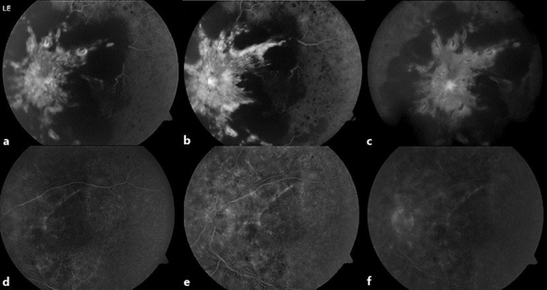
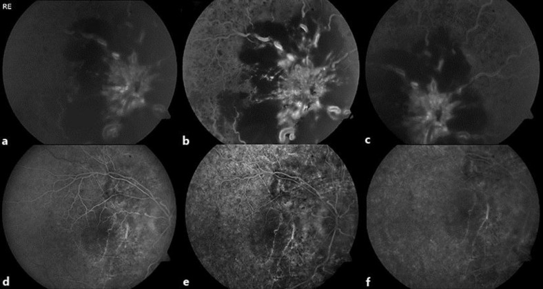
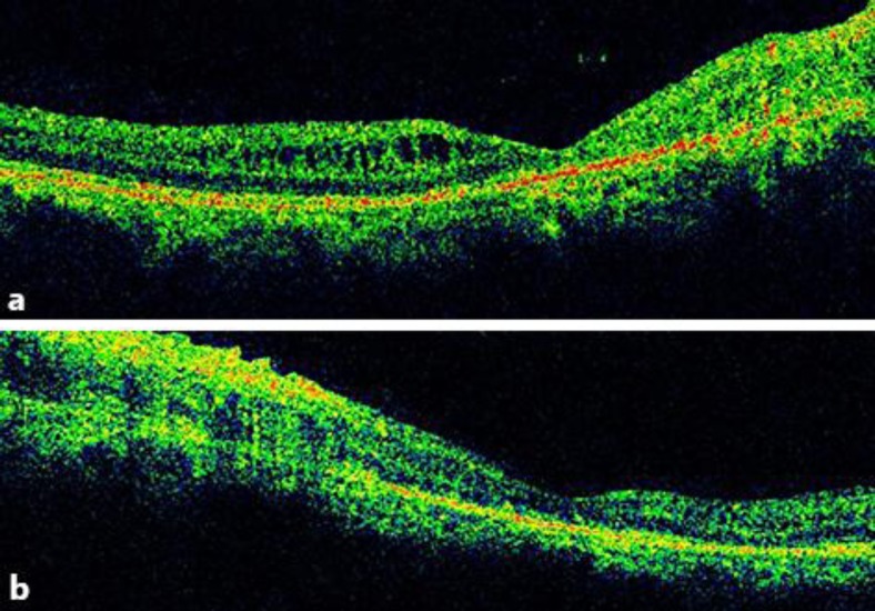
Case report: A 38-year-old man with morbid obesity and daytime sleepiness presented with a history of bilateral vision loss. His visual acuity (VA) was hand movements, and fundus examination (FE) revealed bilateral central RVO. General medical examination revealed untreated hypertension and type II respiratory failure. Laboratory tests for thrombophilia showed increased hematocrit (59%) and high levels of fibrinogen and C-reactive protein. Other causes of congenital and acquired hypercoagulability were ruled out. Pathologic polysomnography led to the diagnosis of OSAS. The patient was treated with antihypertensive drugs and continuous positive air pressure. In addition, he received intravitreal ranibizumab. At 10 months after presentation, his VA was no light perception in the right eye and hand movements in the left eye. FE revealed bilateral retinal and optic nerve atrophy, and the occurrence of a nonarteritic anterior ischemic neuropathy in the right eye was considered.

Keywords: Central retinal vein occlusion; Obstructive sleep apnea syndrome; Optic nerve atrophy; Visual acuity.


Figures

Fig. 1
Fig. 1
Fundus photographs of the left eye. a At presentation, central RVO with optic nerve head swelling, extensive intra- and preretinal hemorrhages in all four quadrants are shown. The macular region is not evaluable. Tortuosity and dilatation of retinal veins are seen. b At 1 month, partial resolution of the hemorrhages are seen. The macular region is visible, showing edema and extensive hard exudation. Retinal venous tortuosity and dilatation are still present. c At 10 months, retinal hemorrhages and macular edema resolved. Optic disc atrophy is seen.
Fig. 2
Fig. 2
Fundus photographs of the right eye. a At presentation, central RVO with extensive hemorrhages that limit the evaluation of the optic disc head is shown. The macular region is not evaluable. b At 1 month, optic disk swelling is visible. Retinal hemorrhages limit evaluation of the macular region, where a hard exudation is seen. Extensive retinal vascular tortuosity and dilatation is present. Temporally to the optic disc, head collateral vessels can be seen. c At 10 months, retinal hemorrhages and macular edema resolved. Important optic atrophy is noticed.
Fig. 3
Fig. 3
FA of the left eye. a, b At 1 week, early venous phase (a) and mid phase (b): hyperfluorescence of the optic disc with indistinct borders. Extensive hypofluorescent blockage due to retinal hemorrhages limits evaluation of the perfusion status. Macular edema is noticed. No signs of ischemia are observed. c At 1 week, late phase: hyperfluorescence of the optic disc persists. Extensive hypofluorescent blockage. d–f At 10 months, early venous phase (d), mid (e) and late phase (f): no macular edema is seen. Hyperfluorescence due to staining and retinal atrophy are noticed. In the macular region, areas of hypo- and hyperfluorescence due to chronic changes in retinal pigmented epithelium are seen. In the macular and optic nerve regions, neither retinal ischemia nor new vessels are observed.
Fig. 4
Fig. 4
FA of the right eye. a, b At 1 week, early venous phase (a) and mid phase (b): important blockage as a result to retinal hemorrhages limits evaluation of the posterior pole. Optic disk head swelling is seen. Important vascular tortuosity and dilatation is noticed. The macular region and the optic disc are not fully evaluable. c At 1 week, late phase: optic disc swelling persists as well as blockage due to hemorrhages. d–f At 10 months, early venous (d), mid (e) and late phase (f): areas of hypo- and hyperfluorescence due to chronic changes in the retinal pigmented epithelium and retinal atrophy are seen. In the macular and optic nerve regions, neither retinal ischemia nor new vessels are observed.
Fig. 5
Fig. 5
SD-OCT of the right and left eye. a SD-OCT of the right eye at 10 months from presentation: important macular atrophy can be seen. Small remnants of intraretinal fluid not involving the fovea are seen. b SD-OCT of the left eye at 10 months from presentation: similarly to the right eye, macular atrophy is seen. No intra- or subretinal fluid is noticed.

References

    1. Lahey JM, Tunç M, Kearney J, Modlinski B, Koo H, Johnson RN, Tanaka S. Laboratory evaluation of hypercoagulable states in patients with central retinal vein occlusion who are less than 56 years of age. Ophthalmology. 2002;1:126–131. - PubMed
    1. Kuhli-Hattenbach C, Scharrer I, Lüchtenberg M, Hattenbach LO. Coagulation disorders and the risk of retinal vein occlusion. Thromb Haemost. 2010;103:299–305. - PubMed
    1. Chou KT, Huang CC, Tsai DC, Chen YM, Perng DW, Shiao GM, Lee YC, Leu HB. Sleep apnea and risk of retinal vein occlusion: a nationwide population-based study of Taiwanese. Am J Ophthalmol. 2012;154:200–205. - PubMed
    1. Turati M, Velez-Montoya R, Gonzalez-Mijares CC, Perez-Montesinos A, Quiroz-Mercado H, Garcia-Aguirre G. Bilateral central retinal vein occlusion associated with obesity-hypoventilation syndrome (Pickwickian syndrome) Retin Cases Brief Rep. 2009;3:140–143. - PubMed
    1. Glacet-Bernard A, Leroux les Jardins G, Lasry S, Coscas G, Soubrane G, Souied E, Housset B. Obstructive sleep apnea among patients with retinal vein occlusion. Arch Ophthalmol. 2010;128:1533–1538. - PubMed

Publication types