Review
. 2014 Jun 17:5:296.
doi: 10.3389/fmicb.2014.00296. eCollection 2014.

Coronavirus infection, ER stress, apoptosis and innate immunity

Affiliations
Review

Coronavirus infection, ER stress, apoptosis and innate immunity

To S Fung et al. Front Microbiol. .

Abstract

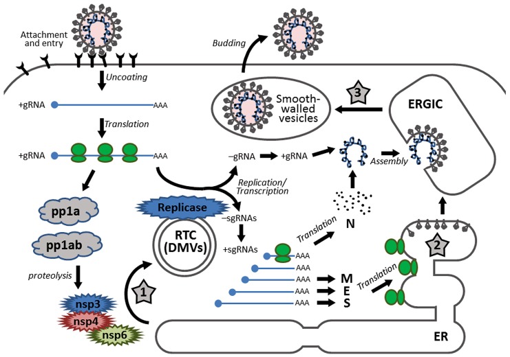
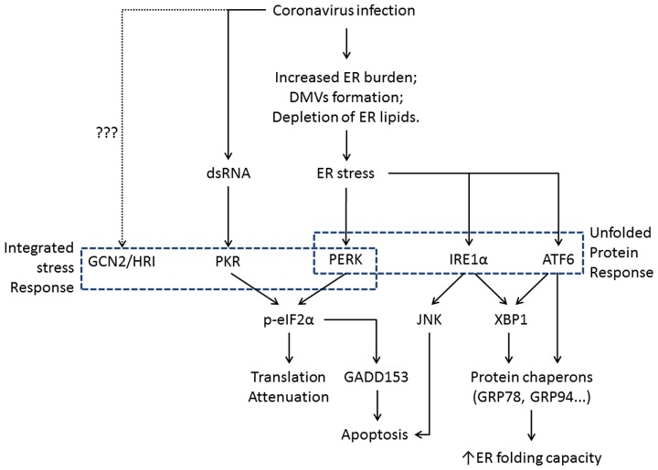
The replication of coronavirus, a family of important animal and human pathogens, is closely associated with the cellular membrane compartments, especially the endoplasmic reticulum (ER). Coronavirus infection of cultured cells was previously shown to cause ER stress and induce the unfolded protein response (UPR), a process that aims to restore the ER homeostasis by global translation shutdown and increasing the ER folding capacity. However, under prolonged ER stress, UPR can also induce apoptotic cell death. Accumulating evidence from recent studies has shown that induction of ER stress and UPR may constitute a major aspect of coronavirus-host interaction. Activation of the three branches of UPR modulates a wide variety of signaling pathways, such as mitogen-activated protein (MAP) kinase activation, autophagy, apoptosis, and innate immune response. ER stress and UPR activation may therefore contribute significantly to the viral replication and pathogenesis during coronavirus infection. In this review, we summarize the current knowledge on coronavirus-induced ER stress and UPR activation, with emphasis on their cross-talking to apoptotic signaling.

Keywords: ER stress; apoptosis; coronavirus; proinflammatory cytokines; signal transduction pathways; unfolded protein response.


Figures

FIGURE 1
FIGURE 1
Schematic diagram showing the replication cycle of coronavirus and the stages in which ER stress may be induced during coronavirus infection. Infection starts with receptor binding and entry by membrane fusion. After uncoating, the genomic RNA is used as a template to synthesize progeny genomes and a nested set of subgenomic RNAs. The replication transcription centers are closely associated with DMVs, which are proposed to be adopted from the modified ER, possibly by the combined activities of non-structural proteins nsp3, nsp4, and nsp6. The S, E, and M proteins are synthesized and anchored on the ER, whereas the N protein is translated in the cytosol. Assembly takes place in the ERGIC and mature virions are released via smooth-walled vesicles by exocytosis. The three stages that presumably induce ER stress are highlighted with numbered star signs, namely: (1) formation of DMVs, (2) massive production and modification of structural proteins, and (3) depletion of ER membrane during budding.
FIGURE 2
FIGURE 2
Flowchart showing the induction of ER stress and its physiological outcomes during coronavirus infection. The integrated stress response pathways (including PERK) trigger translation shutdown and modulate apoptosis. The ATF6 pathway enhances the ER folding capacity, and the IRE1 pathway affects both ER folding and apoptosis induction. Pointed arrows indicate activation. The dotted line suggests uncharacterized function of GCN2 and HRI during coronavirus infection.
FIGURE 3
FIGURE 3
Working model of PKR/PERK-eIF2α-ATF4-GADD153 pathway activation during coronavirus infection, using IBV as an example. Phosphorylation of eIF2α by PERK and PKR induces the expression of ATF4, ATF3, and GADD153. GADD153 exerts its pro-apoptotic activities via suppressing Bcl2 and ERKs by inducing TRIB3. The potential induction of DUSP1 by ATF3 may modulate phosphorylation of p38 and JNK, thus regulating IBV-induced apoptosis and cytokine production. The translation attenuation due to eIF2α activation can also lead to reduced inhibition of IκBα on NF-κB, which in turn promote cytokine production. Pointed arrows indicate activation, and blunt-ended lines indicate inhibition. The question mark indicates hypothetical mechanism.
FIGURE 4
FIGURE 4
Working model of IRE1-XBP1 signaling pathway during coronavirus infection, using IBV as an example. IRE1 mediates XBP1 splicing, which up-regulates UPR target genes to restore ER stress, and the spliced XBP1 may also modulate the IFN and cytokine secretion. IRE1 activation modulates the phosphorylation of Akt and JNK, thus affecting IBV-induced apoptosis. IRE1 is also responsible for basal activity of IKK, which phosphorylates IκBα to remove its inhibition on NF-κB, thus facilitating the production of type I IFN and pro-inflammatory cytokines. Pointed arrows indicate activation, and blunt-ended lines indicate inhibition. The question mark indicates hypothetical mechanism.

References

    1. Alagaili A. N., Briese T., Mishra N., Kapoor V., Sameroff S. C., Burbelo P. D., et al. (2014). Middle East respiratory syndrome coronavirus infection in dromedary camels in Saudi Arabia. MBio 5 e00884–14 10.1128/mBio.01002-14 - DOI - PMC - PubMed
    1. Ambrose R. L., Mackenzie J. M. (2011). West Nile virus differentially modulates the unfolded protein response to facilitate replication and immune evasion. J. Virol. 85 2723–2732 10.1128/JVI.02050-10 - DOI - PMC - PubMed
    1. Ambrose R. L., Mackenzie J. M. (2013). ATF6 signaling is required for efficient West Nile virus replication by promoting cell survival and inhibition of innate immune responses. J. Virol. 87 2206–2214 10.1128/JVI.02097-12 - DOI - PMC - PubMed
    1. Angelini M. M., Akhlaghpour M., Neuman B. W., Buchmeier M. J. (2013). Severe acute espiratory syndrome coronavirus nonstructural proteins 3, 4, and 6 induce double-membrane vesicles. MBio 4:e00524-00513 10.1128/mBio.00524-13 - DOI - PMC - PubMed
    1. Annan A., Baldwin H. J., Corman V. M., Klose S. M., Owusu M., Nkrumah E. E., et al. (2013). Human betacoronavirus 2c EMC/2012-related viruses in bats, Ghana and Europe. Emerg. Infect. Dis. 19 456–459 10.3201/eid1903.121503 - DOI - PMC - PubMed