

Renal infarction in a patient with pulmonary vein thrombosis after left upper lobectomy
- PMID: 24987406
- PMCID: PMC4067727
- DOI: 10.1159/000363224
Renal infarction in a patient with pulmonary vein thrombosis after left upper lobectomy
Abstract
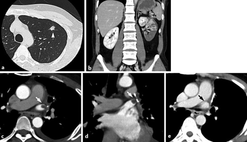
A 43-year-old male experienced renal infarction (RI) following left upper lobectomy for lung cancer. The patient complained of acute-onset severe left flank pain on the 14th postoperative day. A contrast-enhanced computed tomography (CT) of the abdomen revealed RI by a large wedge-shaped defect in the left kidney. A chest CT scan located the thrombus in the stump (a blind-ended vessel) of the left superior pulmonary vein. Therefore, thromboembolic RI caused by pulmonary vein thrombosis was suspected. Anticoagulation therapy was initiated with heparin and warfarin to treat RI and to prevent further embolic episodes. Two months later, pulmonary vein thrombosis had resolved without the appearance of additional peripheral infarction. This case emphasizes the need to consider thrombus in the stump of the pulmonary vein as a cause of RI.
Keywords: Lung cancer; Lung resection; Peripheral infarction; Pulmonary vein thrombosis; Renal infarction.
Figures
References
-
- Bolderman R, Oyen R, Verrijcken A, Knockaert D, Vanderschueren S. Idiopathic renal infarction. Am J Med. 2006;119:356.e9–e12. - PubMed
-
- Antopolsky M, Simanovsky N, Stalnkowicz R, Salameh S, Hiller N. Renal infarction in the ED: 10-year experience and review of the literature. Am J Emerg Med. 2012;30:1055–1060. - PubMed
-
- Bourgault M, Grimbert P, Verret C, Pourrat J, Herody M, Halimi JM, Karras A, Amoura Z, Jourde-Chiche N, Izzedine H, Francois H, Boffa JJ, Hummel A, Bernadet-Monrozies P, Fouque D, Canoui-Poitrine F, Lang P, Daugas E, Audard V. Acute renal infarction: a case series. Clin J Am Soc Nephrol. 2013;8:392–398. - PMC - PubMed
-
- Carey HB, Boltax R, Dickey KW, Finkelstein FO. Bilateral renal infarction secondary to paradoxical embolism. Am J Kidney Dis. 1999;34:752–755. - PubMed
Publication types
LinkOut - more resources
Full Text Sources
Other Literature Sources
