Molecular cloning of a hyaluronidase from Bothrops pauloensis venom gland
- PMID: 24987408
- PMCID: PMC4077683
- DOI: 10.1186/1678-9199-20-25
Molecular cloning of a hyaluronidase from Bothrops pauloensis venom gland
Abstract
Background: Hyaluronate is one of the major components of extracellular matrix from vertebrates whose breakdown is catalyzed by the enzyme hyaluronidase. These enzymes are widely described in snake venoms, in which they facilitate the spreading of the main toxins in the victim's body during the envenoming. Snake venoms also present some variants (hyaluronidases-like substances) that are probably originated by alternative splicing, even though their relevance in envenomation is still under investigation. Hyaluronidases-like proteins have not yet been purified from any snake venom, but the cDNA that encodes these toxins was already identified in snake venom glands by transcriptomic analysis. Herein, we report the cloning and in silico analysis of the first hyaluronidase-like proteins from a Brazilian snake venom.
Methods: The cDNA sequence of hyaluronidase was cloned from the transcriptome of Bothrops pauloensis venom glands. This sequence was submitted to multiple alignment with other related sequences by ClustalW. A phylogenetic analysis was performed using MEGA 4 software by the neighbor joining (NJ) method.
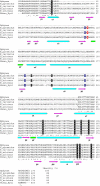
Results: The cDNA from Bothrops pauloensis venom gland that corresponds to hyaluronidase comprises 1175 bp and codifies a protein containing 194 amino acid residues. The sequence, denominated BpHyase, was identified as hyaluronidase-like since it shows high sequence identities (above 83%) with other described snake venom hyaluronidase-like sequences. Hyaluronidases-like proteins are thought to be products of alternative splicing implicated in deletions of central amino acids, including the catalytic residues. Structure-based sequence alignment of BpHyase to human hyaluronidase hHyal-1 demonstrates a loss of some key secondary structures. The phylogenetic analysis indicates an independent evolution of BpHyal when compared to other hyaluronidases. However, these toxins might share a common ancestor, thus suggesting a broad hyaluronidase-like distribution among venomous snakes.
Conclusions: This work is the first report of a cDNA sequence of hyaluronidase from Brazilian snake venoms. Moreover, the in silico analysis of its deduced amino acid sequence opens new perspectives about the biological function of hyaluronidases-like proteins and may direct further studies comprising their isolation and/or recombinant production, as well as their structural and functional characterization.
Keywords: Alternative splicing; Hyaluronidase-like; Snake venom.
Figures


References
-
- Meyer K. In: The Enzymes. Volume 5. Boyer PD, editor. New York: Academic Press; 1971. Hyaluronidases; pp. 307–320.
-
- Menzel EJ, Farr C. Hyaluronidase and its substrate: biochemistry, biological activities and therapeutic uses. Cancer Lett. 1998;131(1):3–11. - PubMed
-
- Csóka AB, Scherer SW, Stern R. Expression analysis of six paralogous human hyaluronidase genes clustered on chromosomes 3p21 and 7q31. Genomics. 1999;60(3):356–361. - PubMed
-
- Duran-Reynalds F. Exaltation de l’activité de virus vaccinal par les extraits de certains organs. Compt Rend Soc Biol. 1928;9:6–7.
LinkOut - more resources
Full Text Sources
Other Literature Sources

