Metastatic mesothelioma to the thyroid
- PMID: 24987442
- PMCID: PMC4058903
- DOI: 10.4103/1742-6413.132984
Metastatic mesothelioma to the thyroid
Abstract
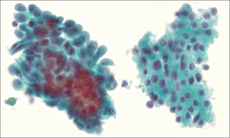
A 69 year-old male patient with a history of malignant mesothelioma treated with chemotherapy and surgical resection with removal of the right lung and right pleural pneumonectomy was clinically in remission for 1 ½ years. A positron emission tomography (PET) scan revealed limited uptake in the right pleural space, thought to represent post-surgical changes, and uptake in the left thyroid lobe. Thyroid ultrasound revealed a solid left lobe nodule with peripheral vascularity and absent microcalcifications. Fine needle aspiration cytology showed a microfollicular arrangement of cytologically bland cells with variable Hürthle cell changes initially interpreted as suspicious for Hürthle cell neoplasm. Review at multidisciplinary conference raised the possibility of metastatic mesothelioma, supported by immunohistochemical studies in the cell block. The patient opted for left hemithyroidectomy with isthmusectomy which confirmed malignant mesothelioma. Repeat PET scan 6 months later revealed no further uptake in the thyroid bed, with limited uptake in the right pleural space. Metastatic tumors to the thyroid are uncommon with only one previous description of metastasis to the thyroid by mesothelioma. Metastasis of cytologically low grade tumors such as mesothelioma present problems for cytology due to the potential for overlap with the variable appearances of thyroid neoplasms. The value (if any) of ancillary tests, including mutation testing, expression profiling and immunohistochemistry is discussed.
Keywords: Nuclear medicine-imaging; pathology-thyroid cytology; thyroid cancer-clinical; thyroid cancer-genetics.
Figures


References
-
- Hegedüs L. Clinical practice.The thyroid nodule. N Engl J Med. 2004;351:1764–71. - PubMed
-
- Soelberg KK, Bonnema SJ, Brix TH, Hegedüs L. Risk of malignancy in thyroid incidentalomas detected by 18F-fluorodeoxyglucose positron emission tomography: A systematic review. Thyroid. 2012;22:918–25. - PubMed
-
- Schmid KW, Hittmair A, Ofner C, Tötsch M, Ladurner D. Metastatic tumors in fine needle aspiration biopsy of the thyroid. Acta Cytol. 1991;35:722–4. - PubMed
-
- Michelow PM, Leiman G. Metastases to the thyroid gland: Diagnosis by aspiration cytology. Diagn Cytopathol. 1995;13:209–13. - PubMed
-
- Ohori NP, Nikiforova MN, Schoedel KE, LeBeau SO, Hodak SP, Seethala RR, et al. Contribution of molecular testing to thyroid fine-needle aspiration cytology of “follicular lesion of undetermined significance/atypia of undetermined significance". Cancer Cytopathol. 2010;118:17–23. - PubMed
Publication types
LinkOut - more resources
Full Text Sources
Other Literature Sources