Review
. 2014 Jun 11;6(3):33.
doi: 10.1186/alzrt263. eCollection 2014.

Cerebral microbleeds: overview and implications in cognitive impairment

Affiliations
Review

Cerebral microbleeds: overview and implications in cognitive impairment

Sergi Martinez-Ramirez et al. Alzheimers Res Ther. .

Abstract

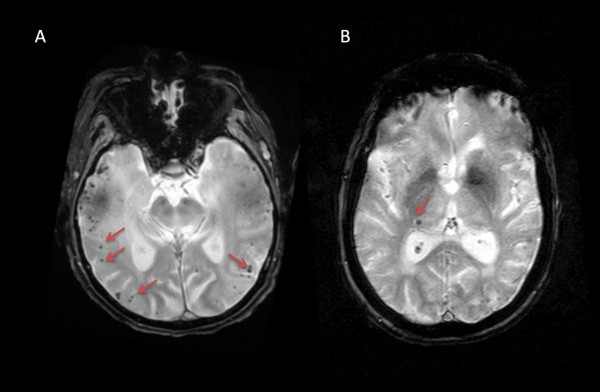
Cerebral microbleeds (MBs) are small chronic brain hemorrhages which are likely caused by structural abnormalities of the small vessels of the brain. Owing to the paramagnetic properties of blood degradation products, MBs can be detected in vivo by using specific magnetic resonance imaging (MRI) sequences. Over the last decades, the implementation of these MRI sequences in both epidemiological and clinical studies has revealed MBs as a common finding in many different populations, including healthy individuals. Also, the topographic distribution of these MBs has been shown to be potentially associated with specific underlying vasculopathies. However, the clinical and prognostic significance of these small hemorrhages is still a matter of debate as well as a focus of extensive research. In this article, we aim to review the current knowledge on the pathophysiology and clinical implications of MBs, with special emphasis on the links between lobar MBs, cerebral amyloid angiopathy, and Alzheimer's disease.


Figures

Figure 1
Figure 1
Cerebral microbleeds as seen on magnetic resonance imaging gradient-recalled echo imaging (arrows). (A) Multiple lobar microbleeds distributed across the temporal lobes. (B) Isolated deep microbleed in the lateral aspect of the right thalamus.

References

    1. Roberts TP, Mikulis D. Neuro MR: principles. J Magn Reson Imaging. 2007;26:823–837. doi: 10.1002/jmri.21029. - DOI - PubMed
    1. Offenbacher H, Fazekas F, Schmidt R, Koch M, Fazekas G, Kapeller P. MR of cerebral abnormalities concomitant with primary intracerebral hematomas. AJNR Am J Neuroradiol. 1996;17:573–578. - PMC - PubMed
    1. Vernooij MW, Ikram MA, Wielopolski PA, Krestin GP, Breteler MM, van der Lugt A. Cerebral microbleeds: accelerated 3D T2*-weighted GRE MR imaging versus conventional 2D T2*-weighted GRE MR imaging for detection. Radiology. 2008;248:272–277. doi: 10.1148/radiol.2481071158. - DOI - PubMed
    1. Haacke EM, Xu Y, Cheng YC, Reichenbach JR. Susceptibility weighted imaging (SWI) Magn Reson Med. 2004;52:612–618. doi: 10.1002/mrm.20198. - DOI - PubMed
    1. Nandigam RN, Viswanathan A, Delgado P, Skehan ME, Smith EE, Rosand J, Greenberg SM, Dickerson BC. MR imaging detection of cerebral microbleeds: effect of susceptibility-weighted imaging, section thickness, and field strength. AJNR Am J Neuroradiol. 2009;30:338–343. - PMC - PubMed

LinkOut - more resources