3D modeling, custom implants and its future perspectives in craniofacial surgery
- PMID: 24987592
- PMCID: PMC4073471
- DOI: 10.4103/2231-0746.133065
3D modeling, custom implants and its future perspectives in craniofacial surgery
Abstract
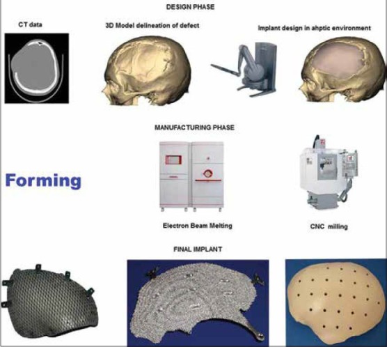
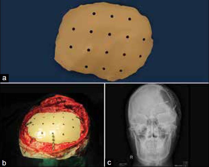
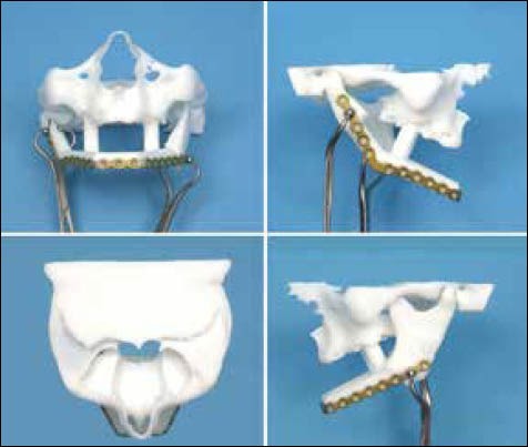
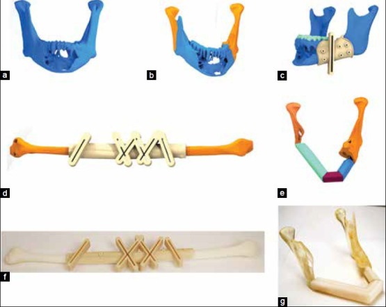
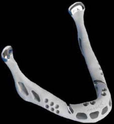
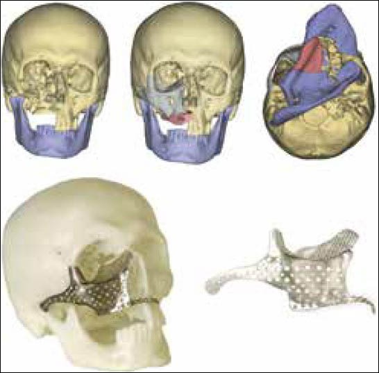
Custom implants for the reconstruction of craniofacial defects have gained importance due to better performance over their generic counterparts. This is due to the precise adaptation to the region of implantation, reduced surgical times and better cosmesis. Application of 3D modeling in craniofacial surgery is changing the way surgeons are planning surgeries and graphic designers are designing custom implants. Advances in manufacturing processes and ushering of additive manufacturing for direct production of implants has eliminated the constraints of shape, size and internal structure and mechanical properties making it possible for the fabrication of implants that conform to the physical and mechanical requirements of the region of implantation. This article will review recent trends in 3D modeling and custom implants in craniofacial reconstruction.
Keywords: 3D modeling; CAD CAM implants; CAD CAM surgery; PEEK implants; additive manufacturing; craniofacial surgery; custom implants; electron beam melting; implants; patient specific implants; porous titanium.
Conflict of interest statement
Figures


References
-
- Shimko DA, Nauman EA. Development and characterization of a porous poly (methyl methacrylate) scaffold with controllable modulus and permeability. J Biomed Mater Res B Appl Biomater. 2007;80:360–9. - PubMed
-
- Schlickewei W, Schlickewei C. The use of bone substitutes in the treatment of bone defects-The clinical view and history. Macromol Symp. 2007;253:10–23.
-
- Lane JM, Sandhu HS. Current approaches to experimental bone grafting. Orthop Clin North Am. 1987;18:213–25. - PubMed
-
- St John TA, Vaccaro AR, Sah AP, Schaefer M, Berta SC, Albert T, et al. Physical and monetary costs associated with autogenous bone graft harvesting. Am J Orthop (Belle Mead NJ) 2003;32:18–23. - PubMed
-
- Silber JS, Anderson DG, Daffner SD, Brislin BT, Leland JM, Hilibrand AS, et al. Donor site morbidity after anterior iliac crest bone harvest for single-level anterior cervical discectomy and fusion. Spine (Phila Pa 1976) 2003;28:134–9. - PubMed
Publication types
LinkOut - more resources
Full Text Sources
Other Literature Sources
Medical
Miscellaneous