. 2014 Jul 2;9(7):e101364.
doi: 10.1371/journal.pone.0101364. eCollection 2014.

Alzheimer's associated β-amyloid protein inhibits influenza A virus and modulates viral interactions with phagocytes

Affiliations

Alzheimer's associated β-amyloid protein inhibits influenza A virus and modulates viral interactions with phagocytes

Mitchell R White et al. PLoS One. .

Abstract

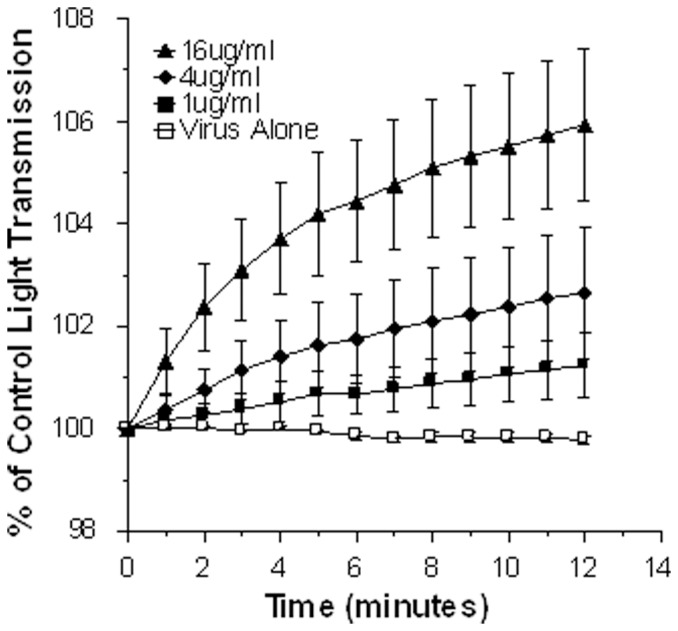
Accumulation of β-Amyloid (βA) is a key pathogenetic factor in Alzheimer's disease; however, the normal function of βA is unknown. Recent studies have shown that βA can inhibit growth of bacteria and fungi. In this paper we show that βA also inhibits replication of seasonal and pandemic strains of H3N2 and H1N1 influenza A virus (IAV) in vitro. The 42 amino acid fragment of βA (βA42) had greater activity than the 40 amino acid fragment. Direct incubation of the virus with βA42 was needed to achieve optimal inhibition. Using quantitative PCR assays βA42 was shown to reduce viral uptake by epithelial cells after 45 minutes and to reduce supernatant virus at 24 hours post infection. βA42 caused aggregation of IAV particles as detected by light transmission assays and electron and confocal microscopy. βA42 did not stimulate neutrophil H2O2 production or extracellular trap formation on its own, but it increased both responses stimulated by IAV. In addition, βA42 increased uptake of IAV by neutrophils. βA42 reduced viral protein synthesis in monocytes and reduced IAV-induced interleukin-6 production by these cells. Hence, we demonstrate for the first time that βA has antiviral activity and modulates viral interactions with phagocytes.


Conflict of interest statement

Competing Interests: Kevan Hartshorn is currently a member of the Editorial Board of PLOS One. This does not alter the authors' adherence to PLOS ONE Editorial policies and criteria.

Figures

Figure 1
Figure 1. Viral neutralizing activity of βA42 and 40 for seasonal H3N2 and pandemic H1N1 strains of IAV.
Aliquots of the Phil82 H3N2 (panel A) or Cal09 H1N1 (panel B) viral strains were incubated with the indicated concentrations of βA42 or 40 or a scrambled version of βA42 and then these samples were used to infect HBTE cell monolayers and tested for infectious foci 7 hrs later using anti-nucleoprotein antibodies and fluorescence detection. * indicates significant reduction in infectivity compared to control (p<0.05; n = 4 experiments) ** indicates p<0.02 compared with βA40, scrambled βA42 and control (ANOVA analysis) # indicates increased infectivity compared to control.
Figure 2
Figure 2. Viral neutralizing activity of βA42 preparations for various IAV strains.
Experiments were carried out as in figure 1 except that MDCK cells were used instead of HTBE cells. Panel A shows inhibition of two H3N2 strains as indicated. Panel B shows inhibition of three H1N1 strains. Panel C shows effects of a different commercially available preparation of βA42 (called βA42 HFIP) against Phil82 and Cal09 strains. Panel D shows the effect of βA42 on infection of A549 cells by Aichi68 IAV and compares the effects of pre-incubating the virus with βA42 as in panel A (“pre-incubate with IAV”) with pre-incubating the cells with βA42. In the latter case, the βA42 was either left in the cell media when virus was added (“no wash”) or washed off prior to adding the virus (“wash”). Although in the “no wash” setting infectivity was significantly reduced this effect was significantly less than when the virus was pre-incubated with βA42 (p<0.01). Finally panel D also shows the effect of pre-incubating cells with IAV at 4°C to allow binding, followed by adding βA42, and increasing the temperature to 37°C (“βA42 at 4°C”). * indicates significant reduction in viral infectivity compared with control (virus alone) (p<0.01). ** indicates where the Aichi68 strain was inhibited significantly more than the Phil82 strain (panel A). Results shown are mean±SEM of 4 or more experiments.
Figure 3
Figure 3. Effects of βA42 on viral internalization and viral replication in MDCK cells as determined by quantitative RT-PCR.
In panels A, Phil82 IAV was pre-incubated with the indicated concentrations of βA42, followed by incubation of these samples for 45 min with MDCK cells as in figure 1. The cell supernatant and cell pellets were then harvested and viral RNA was extracted followed by PCR assay to determine the amount of virus present in cells and supernatant. βA42 significantly reduced the amount of virus taken up by cells after 45 min of infection. There was a trend to increased virus in the supernatant after 45 min of infection but this was not significant. Panel B shows the results of cell and supernatant assays after 24 hrs of infection. βA42 significantly reduced the amount of virus in both cell and supernatant at this time. * indicates p<0.05 compared with control. Results are mean±SEM of three experiments.
Figure 4
Figure 4. Viral aggregation induced by βA42.
Aggregation of the Aichi68 H3N2 viral strain was assessed based on increased light absorbance at 350(n = 5; p<0.02) at the 16 µg/ml concentration of βA42 as compared to light transmission through the control virus preparation.
Figure 5
Figure 5. Confocal and electron microscopy demonstrating viral aggregates formed by βA42.
Alexafluor 594-labeled Aichi68 IAV was incubated with control buffer or βA42 at the indicated concentrations. After this the virus +/- βA42 samples were added to MDCK cell monolayers as described in Methods. The virus appears red, cell nuclei were stained with Dapi 350 and appear blue, and cell membranes were labeled with WGA-Oregon Green 488. The upper pictures show the same fields as those below but the middle pictures only show the wavelength of virus. Aggregates were also observed on electron microscopy (bottom row of pictures). Results are representative of three similar experiments. The pictures were taken at 100× magnification.
Figure 6
Figure 6. Neutrophil H2O2 responses to IAV alone or IAV treated with βA42 or βA40.
Human neutrophils were treated with buffer alone (PBS), IAV alone (Phil82) or IAV that had been pre-treated with βA42 or scrambled βA42 (panel A) or βA40 (panel B) (all at 16 µg/ml). βA42 caused a significant increase in H2O2 response compared to IAV alone or IAV pre-treated with the scrambled peptide of βA40 (indicated by **). Note that the latter two proteins did not increase the response compared to IAV alone. Results are mean±SEM from 5 experiments (each with a separate neutrophil donor).
Figure 7
Figure 7. βA42 increases neutrophil uptake of IAV and IAV-induced NET formation.
In panel A, neutrophils were treated with FITC-labeled Phil82 IAV alone or virus pre-incubated with the indicated peptides. βA42 and βA42 HFIP significantly increased viral uptake by the neutrophils (indicated by * for both; n = 4; p<0.03 compared with control). The amounts of βA shown are those that were initially incubated with IAV prior to a dilution which occurred when these samples were added to the neutrophils. For example where 50 µg/ml is shown in figure 7A or 9B the final concentration with cells was 14 µg/ml. The scrambled peptide and βA40 did not increase viral uptake. Panel B shows NET formation in response to control buffer, IAV alone, βA42 alone or IAV pre-incubated with either 16 or 32 µg/ml of βA42. Sytox green fluorescence was measured by plate reading fluorometer after 30 min of treatment at 37°C. The results shown are mean±SEM of 5 experiments using separate neutrophil donors. The open bars are results of the stimuli in control cells. The black bars are results obtained with cells pre-treated with the WRW4 peptide which inhibits responses mediated by formyl peptide receptors. * indicates p<0.01 vs control buffer. ** indicates p<0.01 vs IAV alone. The WRW4 peptide did not reduce responses to any stimulus.
Figure 8
Figure 8. Fluorescent micrographs of NET formation in response to IAV and/or βA42.
Panel A: Results are representative of 4 experiments using different neutrophil donors. The figures to the left show Sytox green fluorescence and those to the right in this panel show the same fields under phase contrast microscopy to illustrate how many neutrophils were present. These pictures were taken at 40× and enlarged to match the size of the cells in panel B. Panel B are representative of another set of 3 experiments using Phil82 or Aichi68 viral strains as indicated phase and show Sytox green fluorescence taken at 100× magnification.
Figure 9
Figure 9. Effects of βA42 on interactions of IAV with monocytes: viral protein synthesis (panel A), viral uptake (panel B) and virus-induced cytokine generation (panel C).
Adherent peripheral blood monocytes were infected with Phil82 IAV alone or Phil82 IAV treated with the indicated doses of βA42. In panel A the presence of viral nucleoprotein in the cells was tested as in figure 1 after 24 hours of incubation. * indicates significant reduction compared to virus alone (p<0.03; n = 4 separate blood donors). In panel B the ability of monocytes to take up fluorescently labeled IAV after a 45 min incubation was tested. Increased uptake (p<0.05; n = 4) is indicated by *. In panel C the monocytes were incubated with IAV+/−peptide for 24 hrs and then TNF or IL-6 release into the supernatant was measured by ELISA. IAV alone stimulated increased cytokine production by the cells (see Results section). IL-6 production was significantly reduced by pre-incubation of the virus with βA42 (p<0.02; n = 4; indicated by *). TNF production was not reduced by βA42 (n = 8).

References

    1. Dahlgren KN, Manelli AM, Stine WB Jr, Baker LK, Krafft GA, et al. (2002) Oligomeric and fibrillar species of amyloid-beta peptides differentially affect neuronal viability. J Biol Chem 277: 32046–32053. - PubMed
    1. Masters CL, Selkoe DJ (2012) Biochemistry of amyloid beta-protein and amyloid deposits in Alzheimer disease. Cold Spring Harb Perspect Med 2: a006262. - PMC - PubMed
    1. Selkoe DJ (2008) Biochemistry and molecular biology of amyloid beta-protein and the mechanism of Alzheimer's disease. Handb Clin Neurol 89: 245–260. - PubMed
    1. Jang H, Ma B, Lal R, Nussinov R (2008) Models of toxic beta-sheet channels of protegrin-1 suggest a common subunit organization motif shared with toxic alzheimer beta-amyloid ion channels. Biophys J 95: 4631–4642. - PMC - PubMed
    1. Papareddy P, Morgelin M, Walse B, Schmidtchen A, Malmsten M (2012) Antimicrobial activity of peptides derived from human ss-amyloid precursor protein. J Pept Sci 18: 183–191. - PubMed

Publication types

MeSH terms