. 2014 Jul 29;53(29):4869-79.
doi: 10.1021/bi500467h. Epub 2014 Jul 14.

Investigation of ion binding in chlorite dismutases by means of molecular dynamics simulations

Affiliations

Investigation of ion binding in chlorite dismutases by means of molecular dynamics simulations

Axel Sündermann et al. Biochemistry. .

Abstract

Chlorite dismutases are prokaryotic heme b oxidoreductases that convert chlorite to chloride and dioxygen. It has been postulated that during turnover hypochlorite is formed transiently, which might be responsible for the observed irreversible inactivation of these iron proteins. The only charged distal residue in the heme cavity is a conserved and mobile arginine, but its role in catalysis and inactivation is not fully understood. In the present study, the pentameric chlorite dismutase (Cld) from the bacterium Candidatus Nitrospira defluvii was probed for binding of the low spin ligand cyanide, the substrate chlorite, and the intermediate hypochlorite. Simulations were performed with the enzyme in the ferrous, ferric, and compound I state. Additionally, the variant R173A was studied. We report the parametrization for the GROMOS force field of the anions ClO(-), ClO2(-), ClO3(-), and ClO4(-) and describe spontaneous binding, unbinding, and rebinding events of chlorite and hypochlorite, as well as the dynamics of the conformations of Arg173 during simulations. The findings suggest that (i) chlorite binding to ferric NdCld occurs spontaneously and (ii) that Arg173 is important for recognition and to impair hypochlorite leakage from the reaction sphere. The simulation data is discussed in comparison with experimental data on catalysis and inhibition of chlorite dismutase.


Figures
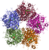
Figure 1
Figure 1
Structure of the pentameric chlorite dismutase from Candidatus Nitrospira defluvii. The five different monomers are colored differently, and the secondary structure elements are shown in a cartoon representation, while the side chains are shown as sticks. The heme is also shown with a stick representation, and the substrate ions in the active sites are shown with a bubble representation.
Figure 2
Figure 2
Normalized distributions of the distance between the heme iron and the Cζ atom of R173. The position of R173 is either “in”, pointing toward the heme iron, or “out” pointing toward the substrate channel entry. The arginine was considered as “in” when then heme iron arginine distance was 0.64 nm or less. This threshold is represented by a vertical black bar in all graphs. Tile A shows the simulation with compound I, hypochlorite, and the arginine starting from an “out” position (CC). Tile B shows the simulation with compound I, hypochlorite, and the arginine starting from an “in” position (CI). Tiles C, E, and G show the simulations with ferric NdCld heme and chlorite, cyanide, and no ion, respectively (OC, ON, OX). Tiles D, F, and H show the simulations with ferrous NdCld heme and chlorite, cyanide, and no ion, respectively (RC, RN, RX). Tiles I and J show the simulations with 20 chlorite ions free in solution with the ferric and ferrous NdCld, respectively (OF, RF).
Figure 3
Figure 3
Normalized distributions of the position of the substrate ion. The substrate ion was considered inside the active site when the distance between the substrate ion and the heme iron was 0.8 nm or less. This threshold is shown by a vertical black bar in all graphs. Tiles A and B show the simulations with the R173A mutant structure and compound I and hypochlorite (MC) and ferric state and chlorite (MO), respectively. Tiles C and D show the simulations with compound I and hypochlorite with the conserved arginine 173 pointing toward the substrate channel entry (CC) or toward the heme iron (CI), respectively. Tiles E and G show the simulations with Fe(III) state and chlorite (OC) and cyanide (ON), respectively. Tiles F and H show the simulations with Fe(II) state and chlorite (RC) and cyanide (RN), respectively. Tiles I and J show the simulations with chlorite ions free in solution and ferric (OF) and ferrous (RF) NdCld, respectively.
Figure 4
Figure 4
Distance between the substrate ions and the heme iron over the course of the simulations. The horizontal black line at 0.8 nm represents the threshold for considering an ion as being inside the active site or outside. Tile A shows the simulation with 20 chlorites free in solution and ferric NdclD (OF). Tile B shows the simulation with 20 chlorites free in solution and ferrous NdCld (RF). Tiles C and D show the simulations with compound I and hypochlorite and the conserved arginine 173 pointing toward the heme iron (CI) and toward the substrate channel entry (CC), respectively. For clarity, only curves for ions that show spontaneous binding are drawn in different colors. When two curves with matching colors are given, these are representative of a single ion interacting with multiple active sites.
Figure 5
Figure 5
Overlay of one active site of the pentameric chlorite dismutase from Candidatus Nitrospira defluvii (NdCld) in the crystal structure with a chlorite bound (in blue) and after spontaneous binding during the simulation with 20 chlorite ions free in solution and an oxidized heme iron (in red). The R173 is pointing toward the substrate channel entry in both cases.
Figure 6
Figure 6
Proposed reaction cycle for chlorite dismutases. First (I) the substrate ClO2 binds to ferric NdCld forming the Fe(III)–ClO2 adduct. R173 possibly plays a role in the initial recognition but is not crucial for the binding. This reaction is followed by oxidation (II) of the heme to compound I with concomitant reduction of chlorite to hypochlorite, ClO. Subsequently, (III) hypochlorite might escape the active site, which is hampered by R173, or (IV) the ferryl oxygen of compound I is rebound by hypochlorite, and (V) chloride and dioxygen are released.

References

    1. Turano P., and Lu Y. (2001) Iron in heme and related proteins, Handbook of metalloproteins, pp 269–356, Marcel Dekker, Inc., New York.
    1. Ebihara A.; Okamoto A.; Kousumi Y.; Yamamoto H.; Masui R.; Ueyama N.; Yokoyama S.; Kuramitsu S. (2005) Structure-based functional identification of a novel heme-binding protein from Thermus thermophilus HB8. J. Struct. Funct. Genomics 6, 21–32. - PubMed
    1. de Geus D. C.; Thomassen E. A.; Hagedoorn P.-L.; Pannu N. S.; van Duijn E.; Abrahams J. P. (2009) Crystal Structure of Chlorite Dismutase, a Detoxifying Enzyme Producing Molecular Oxygen. J. Mol. Biol. 387, 192–206. - PubMed
    1. Goblirsch B. R.; Streit B. R.; DuBois J. L.; Wilmot C. M. (2010) Structural features promoting dioxygen production by Dechloromonas aromatica chlorite dismutase. J. Biol. Inorg. Chem. 15, 879–888. - PMC - PubMed
    1. Kostan J.; Sjöblom B.; Maixner F.; Mlynek G.; Furtmüller P. G.; Obinger C.; Wagner M.; Daims H.; Djinović-Carugo K. (2010) Structural and functional characterisation of the chlorite dismutase from the nitrite-oxidizing bacterium “Candidatus Nitrospira defluvii”: Identification of a catalytically important amino acid residue. J. Struct. Biol. 172, 331–342. - PubMed