. 2014 Jul 2;9(7):e101519.
doi: 10.1371/journal.pone.0101519. eCollection 2014.

The Ran GTPase-activating protein (RanGAP1) is critically involved in smooth muscle cell differentiation, proliferation and migration following vascular injury: implications for neointima formation and restenosis

Affiliations

The Ran GTPase-activating protein (RanGAP1) is critically involved in smooth muscle cell differentiation, proliferation and migration following vascular injury: implications for neointima formation and restenosis

Marc Vorpahl et al. PLoS One. .

Abstract

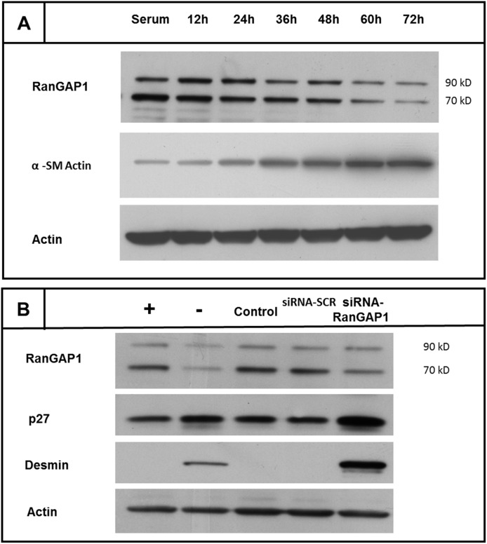
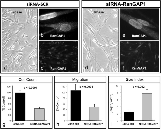
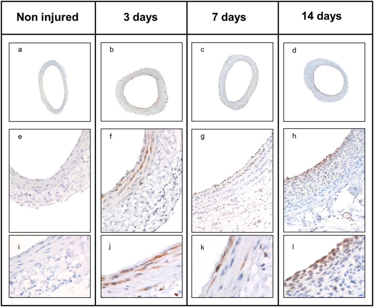
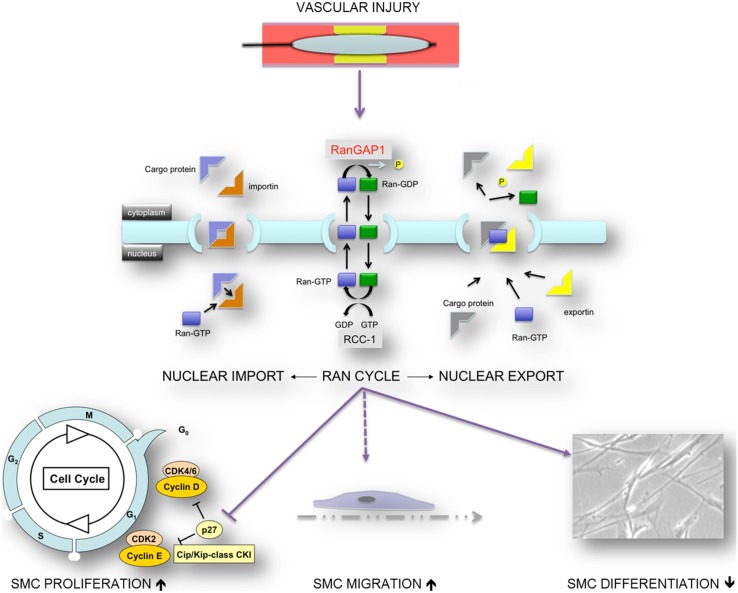
Differentiation and dedifferentiation, accompanied by proliferation play a pivotal role for the phenotypic development of vascular proliferative diseases (VPD), such as restenosis. Increasing evidence points to an essential role of regulated nucleoporin expression in the choice between differentiation and proliferation. However, whether components of the Ran GTPase cycle, which is of pivotal importance for both nucleocytoplasmic transport and for mitotic progression, are subject to similar regulation in VPD is currently unknown. Here, we show that differentiation of human coronary artery smooth muscle cell (CASMC) to a contractile phenotype by stepwise serum depletion leads to significant reduction of RanGAP1 protein levels. The inverse event, dedifferentiation of cells, was assessed in the rat carotid artery balloon injury model, a well-accepted model for neointima formation and restenosis. As revealed by temporospatial analysis of RanGAP1 expression, neointima formation in rat carotid arteries was associated with a significant upregulation of RanGAP1 expression at 3 and 7 days after balloon injury. Of note, neointimal cells located at the luminal surface revealed persistent RanGAP1 expression, as opposed to cells in deeper layers of the neointima where RanGAP1 expression was less or not detectable at all. To gain first evidence for a direct influence of RanGAP1 levels on differentiation, we reduced RanGAP1 in human coronary artery smooth muscle cells by siRNA. Indeed, downregulation of the essential RanGAP1 protein by 50% induced a differentiated, spindle-like smooth muscle cell phenotype, accompanied by an upregulation of the differentiation marker desmin. Reduction of RanGAP1 levels also resulted in a reduction of mitogen induced cellular migration and proliferation as well as a significant upregulation of the cyclin-dependent kinase inhibitor p27KIP1, without evidence for cellular necrosis. These findings suggest that RanGAP1 plays a critical role in smooth muscle cell differentiation, migration and proliferation in vitro and in vivo. Appropriate modulation of RanGAP1 expression may thus be a strategy to modulate VPD development such as restenosis.


Conflict of interest statement

Competing Interests: The authors have declared that no competing interests exist.

Figures

Figure 1
Figure 1. Downregulation of RanGAP1 in arrested, differentiated smooth muscle cells (A).
To induce differentiation, CASMCs were depleted from serum. RanGAP1 expression in CASMC was assessed by Western Blotting 12(∼70 kDa) form as well as the SUMO-1 conjugated (∼90 kDa) form of the RanGAP-1 protein. Both bands revealed reduced RanGAP1 expression over time in cells entering quiescence. α-SM Actin protein, a marker of cell differentiation, is increased over time. Actin levels are displayed as loading control. Effect of posttranscriptional gene silencing of RanGAP1 by small interfering RNA (siRNA) on cell cycle and differentiation markers (B). siRNA mediated gene silencing of RanGAP1 was able to reduce the 90 kD band by 31.8±21.2% (90 kD band) and 75%±14.7% (70 kD band) 48 h post transfection, respectively (average of three different experiments). RanGAP1 depletion was associated with a strong increase of p27Kip1 expression by 60±34%. RanGAP1 deficiency was also associated with a sharp increase in desmin expression with levels even higher than in quiescent cells. CASMC denotes coronary artery smooth muscle cells; (+) denotes serum stimulated CASMC; (−) denotes quiescent CASMC (≥72 h serum depletion); “control” denotes oligofectamine transfected cells without siRNAs; siRNA-SCR denotes scrambled (control) siRNA.
Figure 2
Figure 2. Gene silencing of RanGAP1 by siRNA in CASMC.
CASMC fixed and permeabilized with 4% PFA and 0.2% Triton X-100 were subjected to indirect immunofluorescence with an RanGAP1 antibody. CASMC treated with control scrambled siRNA reveal accumulation of RanGAP1 expression at the nuclear rim rather than in the cytosol (a–c). Likewise, siRNA-RanGAP1 mediated gene silenced CASMC show a residual RanGAP1 expression mainly at the nuclear rim (d–f). Gene silencing of RanGAP1 by means of specific siRNA transfection lead to inhibition of proliferation by 57.4±4.8% (p<0.0001) (g). Similarly, mitogen-induced CASMC migration was sharply inhibited by 48±9% in RanGAP1 siRNA transfected cells (p = 0.0001) (h). Concomitantly, the phenotype of siRNA RanGAP1 treated CASMC showed a significant difference in the cellular size index (length/width; 7.8±2.5 vs. 2.5±0.9 p = 0.002) (i), indicating a phenotypic change that is consistent with contractile, quiescent CASMC.
Figure 3
Figure 3. RanGAP1 expression in the rat carotid artery injury model.
To determine the spatiotemporal expression pattern of RanGAP1 during the course of neointima formation, the rat carotid injury model was applied. Immunohistochemical staining revealed upregulation of RanGAP1 at day 3 (b, f, j, and day 7 (c, g, k) whereas RanGAP1 expression ceased when SMC proliferation decreases at day 14 (d, h, l) subsequent to balloon injury.
Figure 4
Figure 4. Quantitative morphometric and immunohistochemical analysis of neointima formation and RanGAP1 expression in the rat carotid artery injury model.
No significant difference was detectable between the injured group compared to non-injured control arteries with respect to medial area at 3 days, 7 days and 14 days post injury (A) (p = 0.58). Of note, we observed in the media a trend towards a cellular upregulation of RanGAP1 (p = 0.08) 3 days after injury concomitantly with the beginning of cellular proliferation as a response to vascular injury. The increase of neointimal area was detectable at day 7 and peaked at 14 days following vascular injury (B) (p<0.0001). RanGAP1 expression in the media was the highest at the initiation of cellular proliferation and decreased to barely detectable levels at the completion of neointima formation (C). 3 days post injury, almost all cells in the neointima stained positive for RanGAP1 and subsequently, levels decrementally decreased at later time points, e.g. at day 7 and day 14 (D). In non-injured control sections, RanGAP1 expression was virtually undetectable.
Figure 5
Figure 5. Model: RanGAP1, a key player in nucleocytoplasmic transport, plays a critical role in smooth muscle cell differentiation, migration and proliferation.
Appropriate modulation of RanGAP1 expression may thus be a strategy to modulate vascular proliferative disease development such as restenosis.

Similar articles

Cited by

References

    1. Ross R (1999) Atherosclerosis is an inflammatory disease. Am Heart J 138: 419–20. - PubMed
    1. Dzau VJ, Braun-Dullaeus RC, Sedding DG (2002) Vascular proliferation and atherosclerosis: new perspectives and therapeutic strategies. Nat Med 8: 1249–56. - PubMed
    1. Libby P, Aikawa M (2002) Stabilization of atherosclerotic plaques: new mechanisms and clinical targets. Nat Med 8: 1257–62. - PubMed
    1. Libby P, O‘Brien KV (1984) The role of protein breakdown in growth, quiescence and starvation of vascular smooth muscle cells. J Cell Physiol 118: 317–323. - PubMed
    1. Ross R, Glomset JA (1973) Atherosclerosis and the arterial smooth muscle cell: Proliferation of smooth muscle is a key event in the genesis of the lesions of atherosclerosis. Science 180: 1332–9. - PubMed

Substances