. 2014 Jul 1;107(1):88-99.
doi: 10.1016/j.bpj.2014.04.057.

Catch-and-hold activation of muscle acetylcholine receptors having transmitter binding site mutations

Affiliations

Catch-and-hold activation of muscle acetylcholine receptors having transmitter binding site mutations

Prasad Purohit et al. Biophys J. .

Abstract

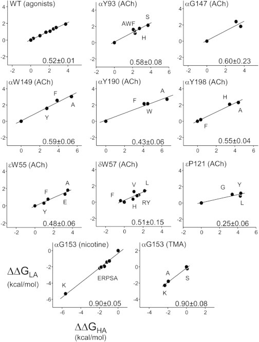
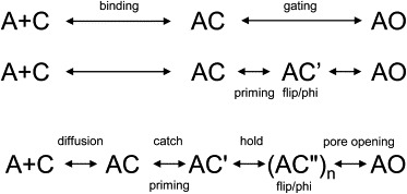
Agonists turn on receptors because their target sites have a higher affinity in the active versus resting conformation of the protein. We used single-channel electrophysiology to measure the lower-affinity (LA) and higher-affinity (HA) equilibrium dissociation constants for acetylcholine in adult-type muscle mouse nicotinic receptors (AChRs) having mutations of agonist binding site amino acids. For a series of agonists and for all mutations of αY93, αG147, αW149, αY190, αY198, εW55, and δW57, the change in LA binding energy was approximately half that in HA binding energy. The results were analyzed as a linear free energy relationship between LA and HA agonist binding, the slope of which (κ) gives the fraction of the overall binding chemical potential where the LA complex is established. The linear correlation between LA and HA binding energies suggests that the overall binding process is by an integrated mechanism (catch-and-hold). For the agonist and the above mutations, κ ∼ 0.5, but side-chain substitutions of two residues had a slope that was significantly higher (0.90; αG153) or lower (0.25; εP121). The results suggest that backbone rearrangements in loop B, loop C, and the non-α surface participate in both LA binding and the LA ↔ HA affinity switch. It appears that all of the intermediate steps in AChR activation comprise a single, energetically coupled process.


Figures

Figure 1
Figure 1
Function and structure. (a) Cyclic model for activation of AChRs having two functionally equivalent agonist sites. LA is lower affinity and HA is higher affinity. C and O are the global closed- and open-channel structural ensembles (LA and HA) and A is the agonist. The bold pathway is Eq. 1. ΔG is the free energy difference between end states. (b) The ligand binding site of the Lymnea acetylcholine binding protein (AChBP; PDB:1UV6 (18)), which is a model for the AChR agonist site. The bound ligand is carbamylcholine (CCh) and the residue numbers are for the mouse α (top) or ε (bottom) subunits. εP121 is a serine in AChBP so only the αC atom is shown. Residues are colored according to their κ-values (see Fig. 5): (purple) 0.90; (green) ∼0.5; (red) 0.25.
Figure 2
Figure 2
Speculative sketch of a single agonist site (Eq. 2). (Top) Cartoon structures. (Arcs) Loops at the agonist site; (solid circle) agonist; (open circles), side chains that stabilize the agonist; (horizontal line at bottom), the conductance of the pore. The left loop rotates in catch and the right in hold. (Middle) Equation 2. A, agonist; C and O, alternative structures of the global transition; superscripts, subconfigurations of the agonist site (′ for catch and ″ for catch+hold; the O state is ″). LA, low affinity; HA, high affinity. Only the first and last steps are by diffusion. Not shown, hold without catch and ligand binding to C′ and C″. (Bottom) Experimental equilibrium constants that pertain to LA binding (1/Kd), gating with one bound agonist (E1), and HA binding (1/Jd).
Figure 3
Figure 3
Using protein engineering methods to estimate equilibrium constants. (a) The un- and diliganded gating equilibrium constants E0 and E2 were estimated from the ratio of open/shut time constants (main component). (Top) Unliganded. Background mutations (αD97A + αY127F + αS269I; no effect on binding) increase spontaneous gating (open is down; the flanks of the cluster are long-lived desensitized states). Adding αY190F reduced E0obs by 1.7-fold. E0WT = 7.4 × 10−7, so with a WT background E0αY190F = 4.3 × 10−7. (Bottom) Diliganded (open is up). Background perturbations (εL269F and depolarization to +100 mV) have no effect on binding and together increase E0 ∼ 15-fold relative to the WT. E2obs = 0.31, so with a WT background (−100 mV) we estimate E2αY190F(ACh) = 0.021. (b) LA association and dissociation rate constants estimated by fitting a sequential model to shut and open interval durations globally, over a range of [ACh]. The apparent equilibrium constants are KdαY190F = 3.6 mM and JdαY190F = 16 μM. Results for other mutations are in Table 1.
Figure 4
Figure 4
Catch-and-hold energy landscape. Energy landscape for agonist binding. The linear correlation of the energies of catch (formation of the LA complex) and catch+hold (the formation of the HA complex) implies that perturbations (agonists or mutations) tilt the overall chemical potential to different extents (dotted lines). We assume that the agonist-site mutations do not alter agonist diffusion or the rest of the events after hold within the global transition. The slope of the plot of ΔGLA versus ΔGHA (κ) gives the position in the overall process at which the LA complex is established. Experimental plots for κ are shown in Fig. 5. For each step, the change in the barrier height is proportional to the change in the end-state energy.
Figure 5
Figure 5
LFERS for agonist binding to adult-type AChRs. The locations of the amino acids are shown in Fig. 1b. The κ-value (linear slope of the ΔΔGLA versus ΔΔGHA plot) is shown at the bottom of each panel. In the top-left panel each symbol is a different agonist and in all other panels each symbol is a different side chain of that position (in all cases, mean of ≥3 patches; Table 1). The WT value is plotted at the origin. A more-positive energy corresponds to a lower affinity.
Figure 6
Figure 6
Nomenclature of intermediate steps and states. (Top) Equation 1: a single intermediate state (AC) between unliganded-C and liganded-O. Binding is by diffusion alone and is independent of the global gating isomerization. (Middle) Two intermediate states (AC and AC′). The superscript indicates a conformational change has occurred at the agonist site. Site-gate communication (priming) occurs upon entry into AC′, which is a nonconducting microstate within the global allosteric transition. (Bottom) Multiple intermediate states, including an encounter complex (AC), an LA complex (AC′), an HA complex, and several more gating microstates ((AC″)n). Superscripts indicate the configuration of the agonist site, the first mark for catch and the second, hold (O is double-prime). Site-gate communication may occur in the catch rearrangement (LA binding).

Comment in

References

    1. Corringer P.J., Le Novère N., Changeux J.P. Nicotinic receptors at the amino acid level. Annu. Rev. Pharmacol. Toxicol. 2000;40:431–458. - PubMed
    1. Nys M., Kesters D., Ulens C. Structural insights into Cys-loop receptor function and ligand recognition. Biochem. Pharmacol. 2013;86:1042–1053. - PubMed
    1. Sine S.M. End-plate acetylcholine receptor: structure, mechanism, pharmacology, and disease. Physiol. Rev. 2012;92:1189–1234. - PMC - PubMed
    1. Unwin N. Nicotinic acetylcholine receptor and the structural basis of neuromuscular transmission: insights from Torpedo postsynaptic membranes. Q. Rev. Biophys. 2013;46:283–322. - PMC - PubMed
    1. Bahar I., Chennubhotla C., Tobi D. Intrinsic dynamics of enzymes in the unbound state and relation to allosteric regulation. Curr. Opin. Struct. Biol. 2007;17:633–640. - PMC - PubMed

Publication types

LinkOut - more resources