. 2014 Jul 2;9(7):e101521.
doi: 10.1371/journal.pone.0101521. eCollection 2014.

The transcription factor MEF2C negatively controls angiogenic sprouting of endothelial cells depending on oxygen

Affiliations

The transcription factor MEF2C negatively controls angiogenic sprouting of endothelial cells depending on oxygen

Caterina Sturtzel et al. PLoS One. .

Abstract

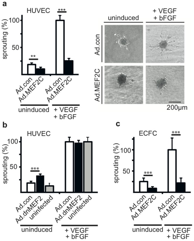
The MADS box transcription factor MEF2C has been detected by us to be upregulated by the angiogenic factors VEGF-A and bFGF in endothelial cells. We have here investigated its potential role for angiogenesis. MEF2C was surprisingly found to strongly inhibit angiogenic sprouting, whereas a dominant negative mutant rather induced sprouting. The factor mainly affected migratory processes of endothelial cells, but not proliferation. In gene profiling experiments we delineated the alpha-2-macroglobulin gene to be highly upregulated by MEF2C. Further data confirmed that MEF2C in endothelial cells indeed induces alpha-2-macroglobulin mRNA as well as the secretion of alpha-2-macroglobulin and that conditioned supernatants of cells overexpressing MEF2C inhibit sprouting. Alpha-2-macroglobulin mediates, at least to a large extent, the inhibitory effects of MEF2C as is shown by knockdown of alpha-2-macroglobulin mRNA by lentiviral shRNA expression which reduces the inhibitory effect. However, under hypoxic conditions the VEGF-A/bFGF-mediated upregulation of MEF2C is reduced and the production of alpha-2-macroglobulin largely abolished. Taken together, this suggests that the MEF2C/alpha-2-macroglobulin axis functions in endothelial cells as a negative feed-back mechanism that adapts sprouting activity to the oxygen concentration thus diminishing inappropriate and excess angiogenesis.


Conflict of interest statement

Competing Interests: The authors have declared that no competing interests exist.

Figures

Figure 1
Figure 1. MEF2C negatively controls angiogenic sprouting.
HUVEC or ECFC were infected with recombinant adenoviruses encoding MEF2C or a dominant negative version of MEF2C (Ad.MEF2C, Ad.dnMEF2C) or empty control viruses (Ad.con) as indicated using MOIs of 3 to 10. Cell spheroids were generated, embedded into collagen gels and induced with VEGF and bFGF (50 ng/ml) or cultured without induction as described in the Methods section. Sprouts were allowed to form for 24 hours. Subsequently pictures were taken for analyses and total sprout length per spheroid assessed using ImageJ software. Data were calculated from a minimum of 15 spheroids per condition and displayed as mean values ± SEM. (a) depicts the effect of MEF2C on HUVEC as calculated from seven independent experiments. The left panel shows the statistical analysis, the right panel exemplary spheroids. (b) shows the effect of a dominant negative version of MEF2 on HUVEC and (c) displays the effect of MEF2C on ECFC as calculated from three independent experiments each. In all parts mean sprout formation induced by VEGF-A and bFGF in Ad.con infected cells is arbitrarily set to 100%. **p<0.005, ***p<0.001.
Figure 2
Figure 2. MEF2C primarily affects migration, but not proliferation, and the effect is independent from Notch signaling and histone acetylase activity.
(a) Inhibition of migration in a wounding assay. A monolayer of HUVEC infected for 48 hours with Ad.MEF2C or Ad.con or left uninfected was scratched and the relative refill of the wounded area was scored and quantified using ImageJ software. The shown results were calculated from three independent experiments performed in triplicates and are displayed as mean values ± SEM. Values obtained from uninfected HUVEC were arbitrarily set to 100%. (b) Absence of a significant effect of MEF2C on actively proliferating cells. The percentage of proliferating cells was assessed for HUVEC seeded in 12 well culture plates and infected with Ad.con, Ad.MEF2C or left uninfected. 24 hours after infection cells were loaded with 2 µM cell proliferation Dye eFlour670, cultured for further 48 hours in complete growth medium and divided cells determined by flow cytometry. Data were calculated from three independent experiments performed in triplicates and displayed as mean values ± SD. (c) Absence of a substantial effect of MEF2C on the increase of cell density. To assess the increase in cell density over a several days period the SRB assay was performed as described in the Methods section. Total protein content was measured after 0, 1, 2 and 3 days of culturing HUVEC infected with Ad.con or Ad.MEF2C in full growth medium in 96 well plates. Data were calculated from three independent experiments performed in quintuplicates and displayed as mean values ± SEM. (d) Effects of inhibition of Notch signaling by DAPT and of histone acetylase activity by TSA. HUVEC were infected with Ad.MEF2C or Ad.con using a MOI of 10. Then they were used in the spheroid sprouting assay stimulated by VEGF and bFGF (50 ng/ml each). Where indicated DAPT (40 µM) or TSA (100 ng/ml) was added. Shown data are mean values ± SD and were calculated from one representative experiment out of three independent experiments performed. Ad.con infected HUVEC induced with VEGF and bFGF but without DAPT or TSA addition were arbitrarily set to 100%. ***p<0.001.
Figure 3
Figure 3. Alpha-2-macroglobulin (A2M) is upregulated by MEF2C in HUVEC on mRNA and protein level.
(a) A2M and MEF2C mRNA levels. HUVEC were transduced for 8, 16, 24 and 32 hours with Ad.MEF2C or Ad.con using MOI of 10 or left without infection (0 hours value). RNAs were isolated from the cells and A2M as well as MEF2C mRNA levels were determined by realtime RT-PCR as described in detail in the Methods section. Fold induction levels are shown as mean values ± SD calculated from triplicates of one representative experiment out of three independent experiments performed. Obtained values were normalized to beta-2-microglobulin mRNA as internal standard. (b) A2M protein. HUVEC were infected with Ad.MEF2C, Ad.con or left without infection for 8 hours, then medium was changed to serum-free Opti-MEM medium and supernatants were harvested after 48 hours. A2M secreted into supernatants was determined using a commercial A2M ELISA kit and a standard curve obtained with purified A2M. The mean concentration of A2M ± SD as calculated from three HUVEC batches analyzed is shown. (c) Supernatants of MEF2C virus transduced HUVEC inhibit sprouting. HUVEC were transduced with Ad.con or Ad.MEF2C or left uninduced for 24 hours, then medium was changed to Opti-mem, after 48 hours supernatants were collected and further concentrated by diafiltration as described in the Methods section. These conditioned media were added to the sprouting assay (50–70 µl to a 500 µl assay) and VEGF-A/bFGF-induced as well as basal sprout formation was scored . Data are displayed as mean values ± SEM as calculated from 3 independent experiments. The values obtained for conditioned medium from Ad.con infected cells treated with VEGF and bFGF were arbitrarily set to 100%. ***p<0.001.
Figure 4
Figure 4. A2M mediates the inhibitive effect of MEF2C on sprouting.
(a) Inhibition of sprouting by purified A2M. Increasing concentrations of commercially available A2M were added to the spheroid sprouting assay and the effects on total sprout length scored. Data are displayed as mean values ± SEM as calculated from three experiments. (b) A2M mRNA expression is strongly reduced upon shRNA mediated knockdown. HUVEC were transduced with LV.shA2M or LV.shcon for 48 hours. Total RNA was isolated, subjected to cDNA synthesis and analyzed by realtime RT-PCR. Values were normalized to beta-2-microglobulin mRNA as internal standard and displayed as mean ±SD. One representative experiment of 3 performed in triplicates is shown (c) Knockdown of A2M restores sprouting activity in Ad.MEF2C transduced HUVEC. Cells were first transduced with LV.shcon or LV.shA2M for 24 hours and then infected with Ad.con or Ad.MEF2C for 6 hours before they were used to form spheroids for the spheroid sprouting assay. Basal and VEGF-A/bFGF-induced sprouting was scored. Data displayed are mean values ± SEM calculated from three experiments. (d) Knockdown of A2M reduces the inhibitory activity in conditioned media from Ad.MEF2C transduced cells. HUVEC were transduced with LV.shcon or LV.shA2M for 24 hours and then infected with Ad.con or Ad.MEF2C for 8 hours. Then medium was changed to serum-free Opti-MEM medium, supernatants were harvested after 48 hours, concentrated by diafiltration and added to the spheroid sprouting assay. Basal and VEGF-A/bFGF-induced sprouting was assessed. Values depicted are mean values ± SEM calculated from three independent experiments. The values obtained for conditioned medium from Ad.con infected cells treated with VEGF and bFGF were arbitrarily set to 100%. n.s. not significant, ***p<0.001.
Figure 5
Figure 5. Under hypoxia the inhibition of sprouting by MEF2C is relieved and VEGF-dependent induction of A2M mRNA is abolished.
(a) Reduction in MEF2C-mediated inhibition of sprouting under hypoxia. HUVEC were transduced with Ad.MEF2C or control adenoviruses and incubated for 48 hours. Then the spheroid sprouting assay was performed without or with addition of VEGF and bFGF as inducers under normoxia or at 1.5% oxygen in the cell culture incubator. 15 spheroids were analyzed per condition. Shown data were calculated from 4 independent experiments and displayed as mean values ± SEM. The values obtained for Ad.con infected cells treated with VEGF and bFGF under normoxia were arbitrarily set to 100%. ∂ indicates difference between hypoxia and normoxia, which is significantly stronger pronounced in Ad.MEF2C transduced HUVEC. **p<0.005, ***p<0.001 (b) Reduction in VEGF-mediated upregulation of MEF2C and A2M mRNA under hypoxia. HUVEC were stimulated with VEGF-A and further incubated under either normoxic or hypoxic conditions for 4, 8, 32 and 56 hours. Total RNA was isolated, subjected to cDNA synthesis and analyzed by realtime RT-PCR. Values were normalized to beta-2-microglobulin mRNA as internal standard and displayed as mean ±SD. One representative experiment of 3 performed in triplicates is shown.

References

    1. Dvorak HF, Brown LF, Detmar M, Dvorak AM (1995) Vascular permeability factor/vascular endothelial growth factor, microvascular hyperpermeability, and angiogenesis. Am J Pathol 146: 1029–1039. - PMC - PubMed
    1. Semenza GL (2003) Targeting HIF-1 for cancer therapy. Nat Rev Cancer 3: 721–732. - PubMed
    1. Phng LK, Gerhardt H (2009) Angiogenesis: a team effort coordinated by notch. Dev Cell 16: 196–208. - PubMed
    1. Hofer E, Schweighofer B (2007) Signal transduction induced in endothelial cells by growth factor receptors involved in angiogenesis. Thromb Haemost 97: 355–363. - PMC - PubMed
    1. Koch S, Tugues S, Li X, Gualandi L, Claesson-Welsh L (2011) Signal transduction by vascular endothelial growth factor receptors. Biochem J 437: 169–183. - PubMed

Publication types

MeSH terms