. 2014 Jul 2;9(7):e100912.
doi: 10.1371/journal.pone.0100912. eCollection 2014.

Staphylococcus aureus sepsis induces early renal mitochondrial DNA repair and mitochondrial biogenesis in mice

Affiliations

Staphylococcus aureus sepsis induces early renal mitochondrial DNA repair and mitochondrial biogenesis in mice

Raquel R Bartz et al. PLoS One. .

Abstract

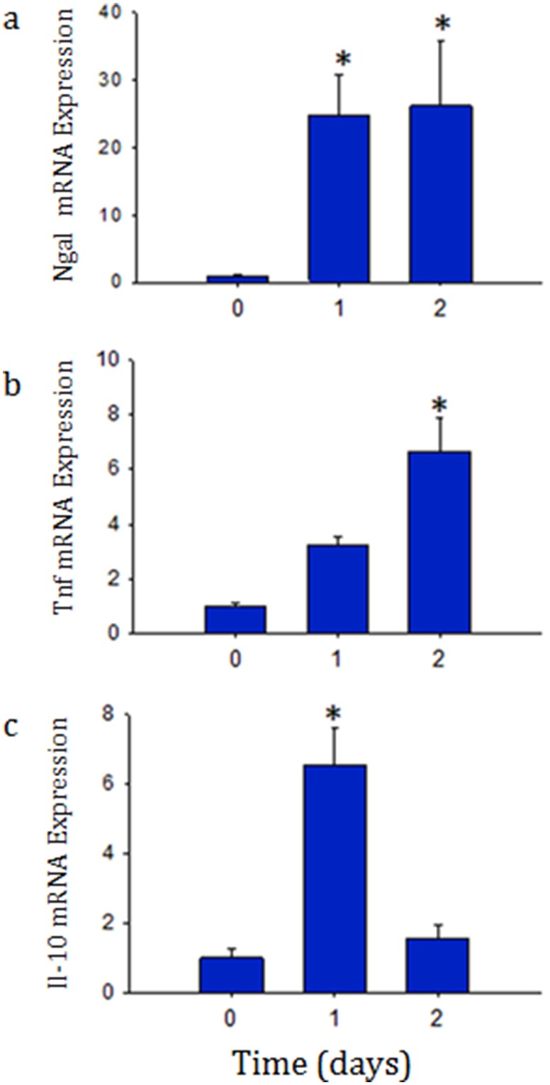
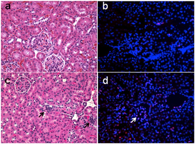
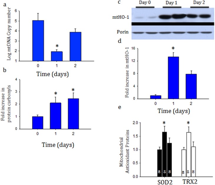
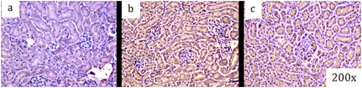
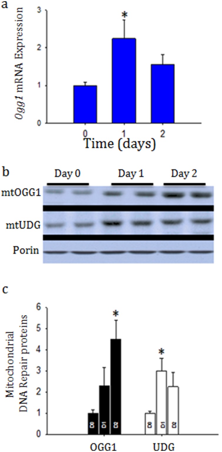
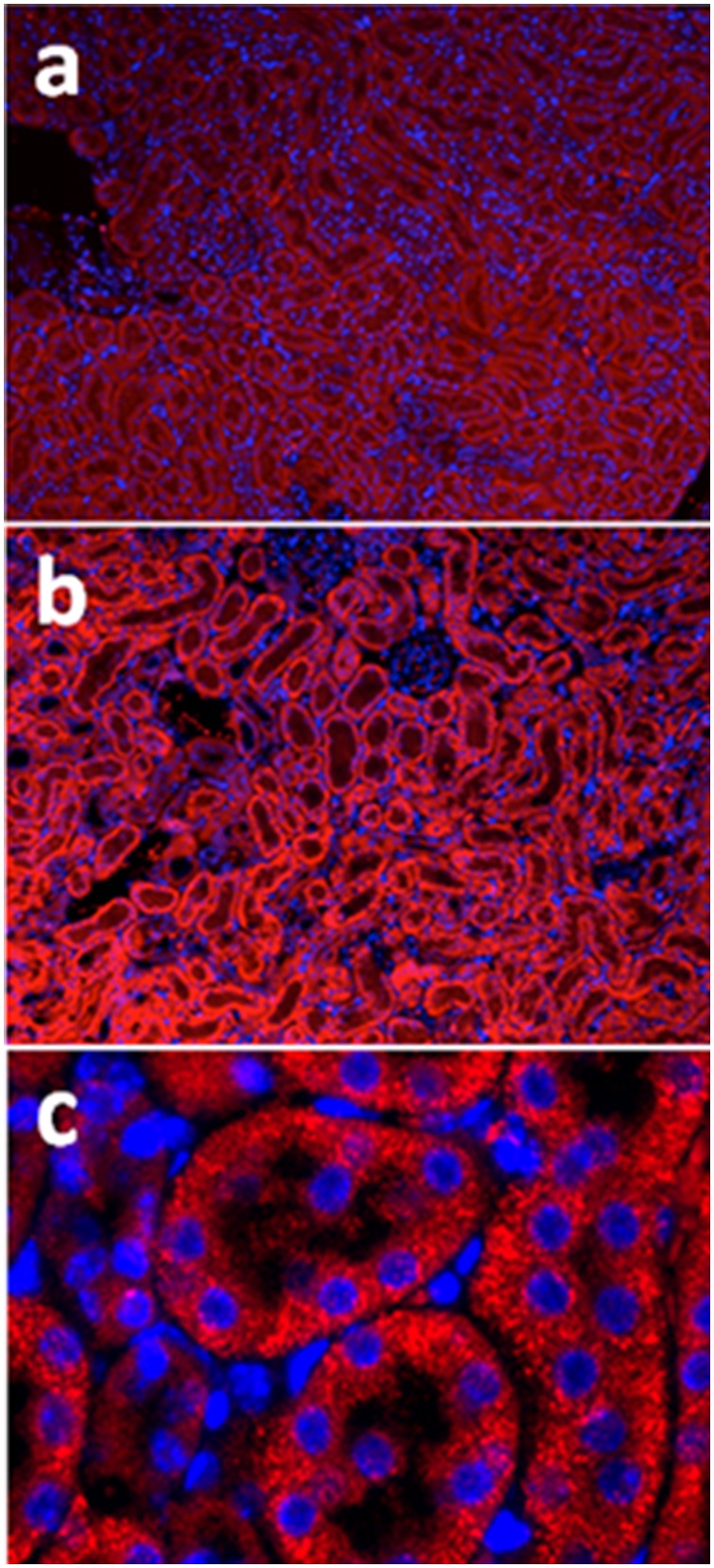
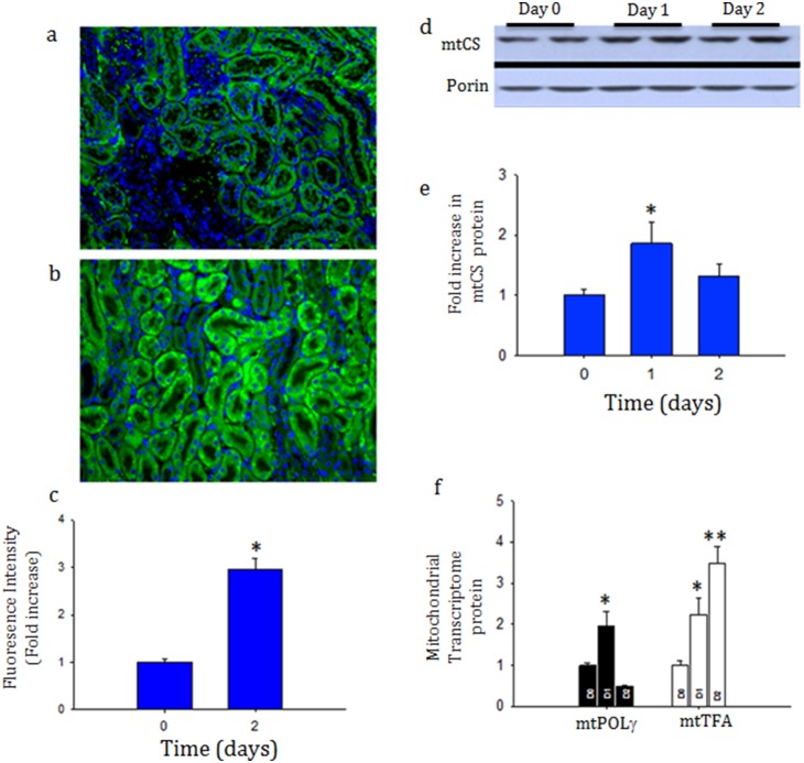
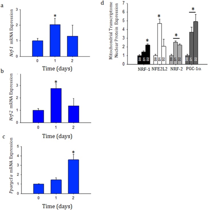
Acute kidney injury (AKI) contributes to the high morbidity and mortality of multi-system organ failure in sepsis. However, recovery of renal function after sepsis-induced AKI suggests active repair of energy-producing pathways. Here, we tested the hypothesis in mice that Staphyloccocus aureus sepsis damages mitochondrial DNA (mtDNA) in the kidney and activates mtDNA repair and mitochondrial biogenesis. Sepsis was induced in wild-type C57Bl/6J and Cox-8 Gfp-tagged mitochondrial-reporter mice via intraperitoneal fibrin clots embedded with S. aureus. Kidneys from surviving mice were harvested at time zero (control), 24, or 48 hours after infection and evaluated for renal inflammation, oxidative stress markers, mtDNA content, and mitochondrial biogenesis markers, and OGG1 and UDG mitochondrial DNA repair enzymes. We examined the kidneys of the mitochondrial reporter mice for changes in staining density and distribution. S. aureus sepsis induced sharp amplification of renal Tnf, Il-10, and Ngal mRNAs with decreased renal mtDNA content and increased tubular and glomerular cell death and accumulation of protein carbonyls and 8-OHdG. Subsequently, mtDNA repair and mitochondrial biogenesis was evidenced by elevated OGG1 levels and significant increases in NRF-1, NRF-2, and mtTFA expression. Overall, renal mitochondrial mass, tracked by citrate synthase mRNA and protein, increased in parallel with changes in mitochondrial GFP-fluorescence especially in proximal tubules in the renal cortex and medulla. Sub-lethal S. aureus sepsis thus induces widespread renal mitochondrial damage that triggers the induction of the renal mtDNA repair protein, OGG1, and mitochondrial biogenesis as a conspicuous resolution mechanism after systemic bacterial infection.


Conflict of interest statement

Competing Interests: The authors have declared that no competing interests exist.

Figures

Figure 1
Figure 1. a, b, and c: Profiling of renal tissue molecular inflammatory markers using Real-time PCR after S. aureus sepsis.
Quantitative real-time PCR for Ngal (a), Tnf (b), and Il-10 (c) mRNA in renal tissue at time zero (control), Day 1 (1), and Day 2 (2) after infection. Ngal mRNA increased more than 20-fold over control by Day 1 of infection, Tnf, and Il-10 also increased significantly, however at different time points. Data represents 6 mice in each group. *P<0.05 is considered significant by ANOVA and Tukey’s post-test analysis.
Figure 2
Figure 2. a, b, c, d: Representative photomicrographs of H&E staining of the renal cortex at time zero (a) and Day 1 (b) and TUNEL staining at time zero (c) and Day 1 after infection (d) after infection.
Arrows indicate areas of necrosis/apoptosis in the H&E stain and TUNEL labeled nuclei. Note inflammatory cell infiltration.
Figure 3
Figure 3. a, b, c, d and e: Mitochondrial DNA copy number based on real-time PCR of cytochrome B/18S levels from kidney (3a) after S. aureus infection.
MtDNA decreased only transiently suggesting active repair and biogenesis. Oxy-blot analysis of whole-cell protein carbonyl content of whole kidney after S. aureus sepsis (3b). Western blot analysis and densitometry of heme-oxygenase 1 (HO-1) levels in the mitochondrial fraction (3c and 3d). Mitochondrial antioxidant enzymes levels of superoxide dismutase (SOD2) and thioredoxin (TRX2) relative to porin densitometry analysis (3e). Data represents 6 mice in each group. *P<0.05 is considered significant by ANOVA and Tukey’s post-test analysis.
Figure 4
Figure 4. a, b, and c: Immunohistochemistry, brown staining, for 8-0H8dG in kidney before (4a), 24 hrs (4b) and 48 hrs (4c) after S. aureus peritoneal sepsis in mice.
Figure 5
Figure 5. a, b, and c: Determination of mitochondrial DNA repair enzymes after sepsis in the crude mitochondrial fraction.
Figure 5a shows quantitative Real-time PCR for Ogg1, a base-excision repair protein especially important for repair of mitochondrial DNA, from whole kidney. Representative western blot analysis for OGG1 and UDG, mitochondrial DNA repair enzymes, compared to Porin from the mitochondrial fraction at control, and at Day 1 and Day 2 after sepsis (5b) and relative densitometry compared from three mice at each time-point (5c). OGG1 increases significantly in the mitochondrial fraction. *P<0.05 significant by ANOVA and Tukey’s post-hoc analysis.
Figure 6
Figure 6. a, b, and c: Representative immunofluorescence for OGG1 (red) in renal tissue before and after infection at time 0 (6a) and Day 2 (6b) after infection at 10x and 40x magnification (6c).
OGG1 increased in the kidneys by immunofluorescence confirming mRNA and Western results.
Figure 7
Figure 7. a, b, c, d, and e: Evidence of mitochondrial biogenesis after sepsis in GFP-tagged mitochondrial Cox8 subunit mice.
Representative immunfluorescence of renal cortex at time zero (7a) and 48 hours (7b) after infection with S. aureus Figure 7c shows the fluorescence intensity of each cortical area. The mitochondrial transcriptome increased after sepsis induction within the mitochondrial compartment as measured mitochondrial citrate synthase (mCS) (7d and 7e) by POLg and mtTFA (7f). Each time-point represents 3 mice. *P<0.05 significant by ANOVA and Tukey’s post-test analysis.
Figure 8
Figure 8. a, b, c, and d: Detection of nuclear markers of mitochondrial biogenesis.
Whole kidney mRNA was evaluated by quantitative real-time PCR for Nrf-1 (8a), Nrf-2 (Gabpa) (8b), and Ppargc1a (8c) at time zero, day 1 and day 2 after infection as well as kidney nuclear protein densitometry for NRF-1, NFE2l2, NRF-2 (GABPa), and PGC-1a relative to histone (8d) by western blot analysis. Each time-point represents 3 mice. *P<0.05 significant by ANOVA and Tukey’s post-test analysis.

References

    1. Uchino S, Kellum JA, Bellomo R, Doig GS, Morimatsu H, et al. (2005) Acute renal failure in critically ill patients: a multinational, multicenter study. JAMA 294: 813–818. - PubMed
    1. Chertow GM, Soroko SH, Paganini EP, Cho KC, Himmelfarb J, et al. (2006) Mortality after acute renal failure: models for prognostic stratification and risk adjustment. Kidney Int 70: 1120–1126. - PubMed
    1. Bagshaw SM, Uchino S, Bellomo R, Morimatsu H, Morgera S, et al. (2007) Septic acute kidney injury in critically ill patients: clinical characteristics and outcomes. Clinical journal of the American Society of Nephrology: CJASN 2: 431–439. - PubMed
    1. Chertow GM, Burdick E, Honour M, Bonventre JV, Bates DW (2005) Acute kidney injury, mortality, length of stay, and costs in hospitalized patients. J Am Soc Nephrol 16: 3365–3370. - PubMed
    1. Langenberg C, Bellomo R, May C, Wan L, Egi M, et al. (2005) Renal blood flow in sepsis. Critical care 9: R363–374. - PMC - PubMed

Publication types

MeSH terms