. 2014 Oct;95(Pt 10):2223-2232.
doi: 10.1099/vir.0.068429-0. Epub 2014 Jul 2.

Consensus proposals for classification of the family Hepeviridae

Affiliations

Consensus proposals for classification of the family Hepeviridae

Donald B Smith et al. J Gen Virol. 2014 Oct.

Erratum in

  • Consensus proposals for classification of the family Hepeviridae.
    Smith DB, Simmonds P, Members Of The International Committee On The Taxonomy Of Viruses Hepeviridae Study Group, Jameel S, Emerson SU, Harrison TJ, Meng XJ, Okamoto H, Van der Poel WHM, Purdy MA. Smith DB, et al. J Gen Virol. 2015 May;96(Pt 5):1191-1192. doi: 10.1099/vir.0.000115. J Gen Virol. 2015. PMID: 26015322 Free PMC article. No abstract available.

Abstract

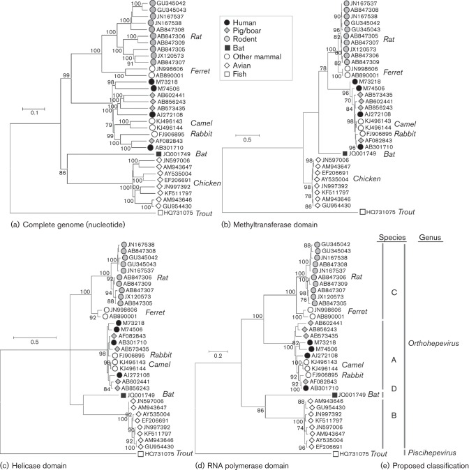
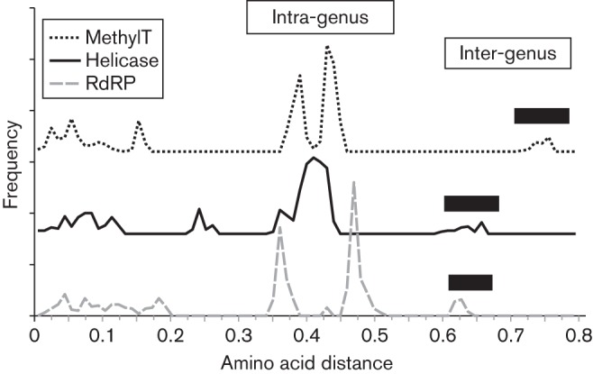
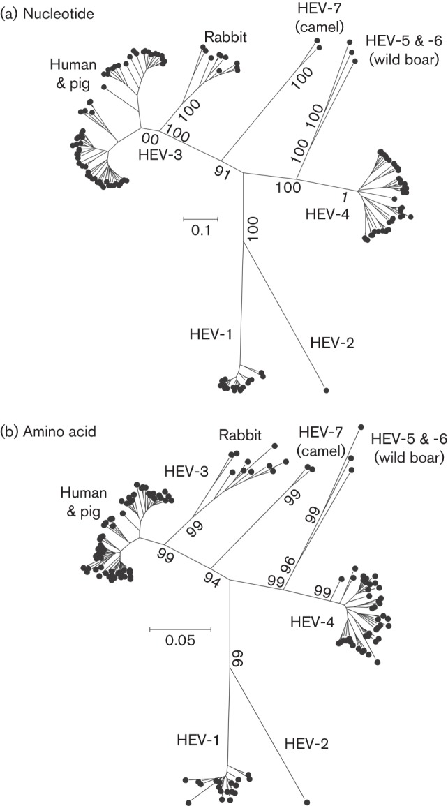
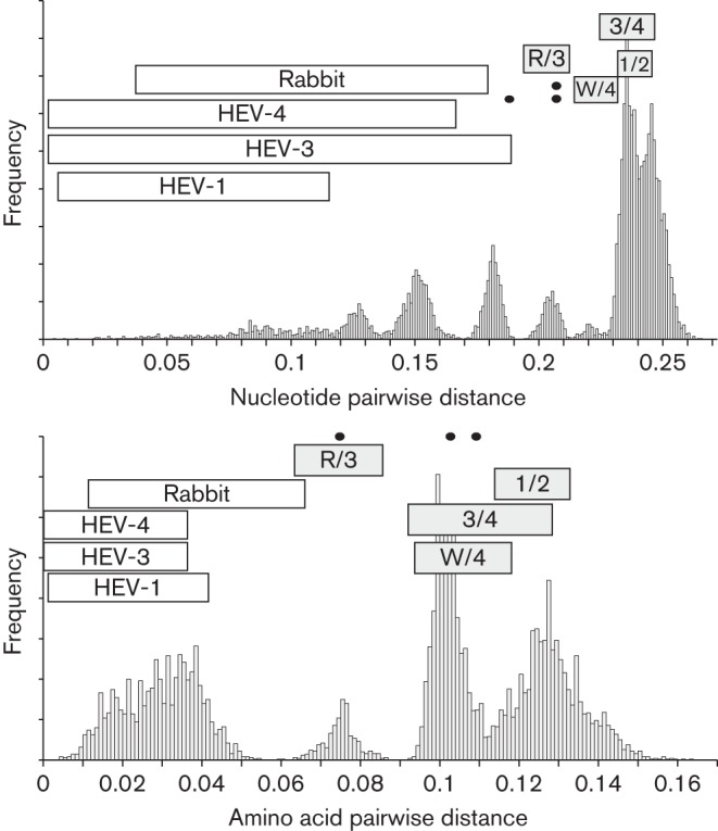
The family Hepeviridae consists of positive-stranded RNA viruses that infect a wide range of mammalian species, as well as chickens and trout. A subset of these viruses infects humans and can cause a self-limiting acute hepatitis that may become chronic in immunosuppressed individuals. Current published descriptions of the taxonomical divisions within the family Hepeviridae are contradictory in relation to the assignment of species and genotypes. Through analysis of existing sequence information, we propose a taxonomic scheme in which the family is divided into the genera Orthohepevirus (all mammalian and avian hepatitis E virus (HEV) isolates) and Piscihepevirus (cutthroat trout virus). Species within the genus Orthohepevirus are designated Orthohepevirus A (isolates from human, pig, wild boar, deer, mongoose, rabbit and camel), Orthohepevirus B (isolates from chicken), Orthohepevirus C (isolates from rat, greater bandicoot, Asian musk shrew, ferret and mink) and Orthohepevirus D (isolates from bat). Proposals are also made for the designation of genotypes within the human and rat HEVs. This hierarchical system is congruent with hepevirus phylogeny, and the three classification levels (genus, species and genotype) are consistent with, and reflect discontinuities in the ranges of pairwise distances between amino acid sequences. Adoption of this system would include the avoidance of host names in taxonomic identifiers and provide a logical framework for the assignment of novel variants.


Figures

Fig. 1.
Fig. 1.
Phylogenetic analysis of members of the family Hepeviridae. (a) Neighbour-joining tree of p-distances among aligned complete genome sequences. (b) Maximum-likelihood tree for amino acid sequences in methyltransferase domain (ORF1-28 to ORF1-389) (c) helicase domain (ORF1-971 to ORF1-1185) (d) RNA-dependent RNA polymerase domain (ORF1-1249 to ORF-1-1671). (e) Proposed classification. Maximum-likelihood trees were computed using the model according to Le & Gascuel with a gamma distribution of evolutionary rates among sites with some invariant sites. Branches supported by >70 % of bootstrap replicates are indicated.
Fig. 2.
Fig. 2.
Frequency distribution of distances among the sequences of members of the family Hepeviridae. Plots show the frequency of amino acid sequence p-distances among amino acid sequences of the methyltransferase (ORF1-28 to ORF1-389, dotted line), helicase (ORF1-971 to ORF1-1185, solid line) and RdRP (ORF1-1249 to ORF1-1671, dashed line) domains. The methyltransferase and helicase distance frequencies are shifted by 160 and 80 respectively in order to improve legibility. Filled bars indicate the range of inter-generic distances.
Fig. 3.
Fig. 3.
Phylogenetic analyses of members of the genus Orthohepevirus. Maximum-likelihood tree for amino acid sequences in (a) ORF1 and (b) ORF2, (c) ORF1-779 to the end of ORF2 with the addition of KF951328 from moose, (d) ORF1-1420 to ORF1-1505 with the addition of sequences from mink, fox, greater bandicoot, Asian musk shrew and bat. Maximum-likelihood trees were computed using the model according to Le & Gascuel with a gamma distribution of evolutionary rates among sites, with some invariant sites. Branches supported by >70 % of bootstrap replicates are indicated.
Fig. 4.
Fig. 4.
Phylogenetic analyses of members of Orthohepevirus A. Maximum-likelihood trees were produced for 137 concatenated ORF1 and ORF2 regions (excluding the HVR and rabbit HEV-specific insertion) for (a) nucleotide sequences using the general time-reversible model with gamma distribution and invariant sites and (b) amino acid sequences using the Jones–Taylor–Thornton model with frequencies, and gamma distribution with invariant sites. Branches supported by >70 % of bootstrap replicates are indicated.
Fig. 5.
Fig. 5.
Frequency distribution of distances among sequences of variants in the genus Orthohepevirus. Histograms show the frequency of nucleotide (a) and amino acid sequence p-distances (b) among 135 concatenated ORF1 and ORF2 sequences (excluding the HVR and rabbit HEV-specific HVR insertion). Maximum and minimum intra-genotype ranges of distances within genotypes 1, 3 and 4 and between rabbit sequences are indicated with open bars. Grey-filled bars depict distances between HEV-3 and rabbit variants (R/3) and inter-genotype distances (3/4, HEV-3 and HEV-4; 1/2, HEV-1 and HEV-2; W/4, wild boar and HEV-4). Distances between the three wild boar sequences are indicated with spots.

Similar articles

Cited by

References

    1. Batts W., Yun S., Hedrick R., Winton J. (2011). A novel member of the family Hepeviridae from cutthroat trout (Oncorhynchus clarkii). Virus Res 158, 116–123 10.1016/j.virusres.2011.03.019 - DOI - PubMed
    1. Bilic I., Jaskulska B., Basic A., Morrow C. J., Hess M. (2009). Sequence analysis and comparison of avian hepatitis E viruses from Australia and Europe indicate the existence of different genotypes. J Gen Virol 90, 863–873 10.1099/vir.0.007179-0 - DOI - PubMed
    1. Bodewes R., van der Giessen J., Haagmans B. L., Osterhaus A. D. M. E., Smits S. L. (2013). Identification of multiple novel viruses, including a parvovirus and a hepevirus, in feces of red foxes. J Virol 87, 7758–7764 10.1128/JVI.00568-13 - DOI - PMC - PubMed
    1. Chen X., Zhang Q., He C., Zhang L., Li J., Zhang W., Cao W., Lv Y.-G., Liu Z. & other authors (2012). Recombination and natural selection in hepatitis E virus genotypes. J Med Virol 84, 1396–1407 10.1002/jmv.23237 - DOI - PubMed
    1. Dai X., Dong C., Zhou Z., Liang J., Dong M., Yang Y., Fu J., Tian H., Wang S. & other authors (2013). Hepatitis E virus genotype 4, Nanjing, China, 2001-2011. Emerg Infect Dis 19, 1528–1530 10.3201/eid1909.130013 - DOI - PMC - PubMed

Publication types