. 2014 Jul 3:5:4286.
doi: 10.1038/ncomms5286.

The fidelity of the ligation step determines how ends are resolved during nonhomologous end joining

Affiliations

The fidelity of the ligation step determines how ends are resolved during nonhomologous end joining

Crystal A Waters et al. Nat Commun. .

Abstract

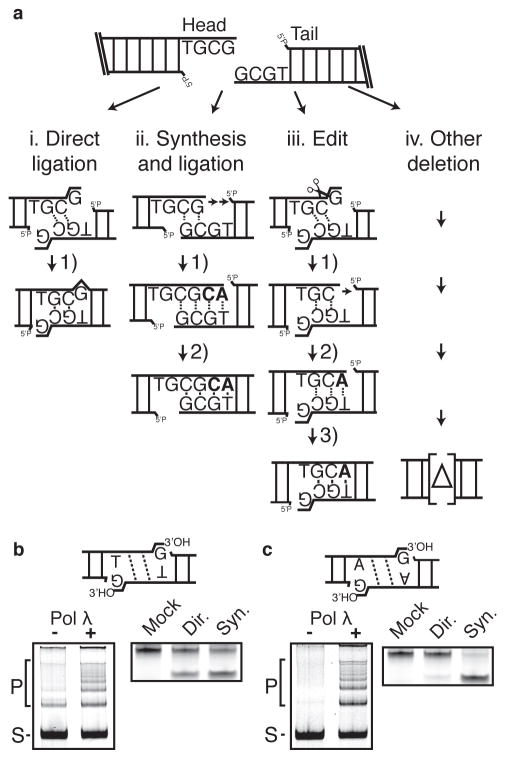
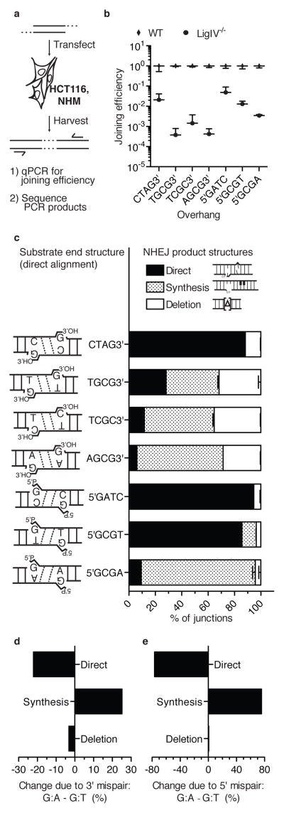
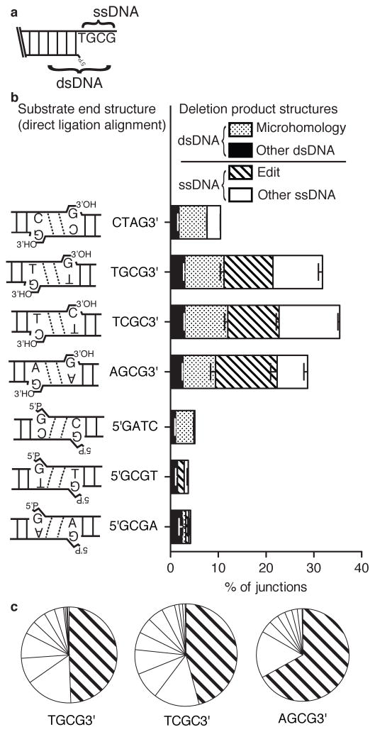
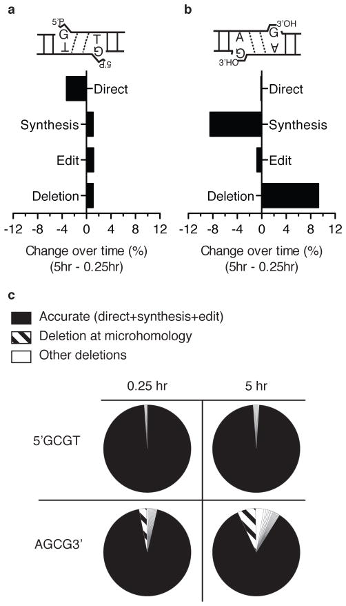
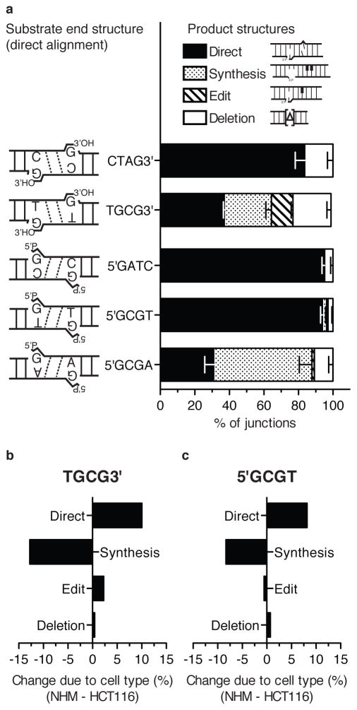
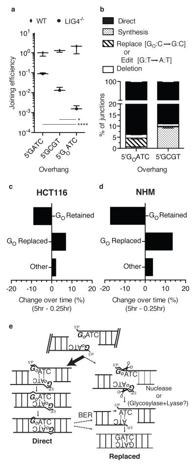
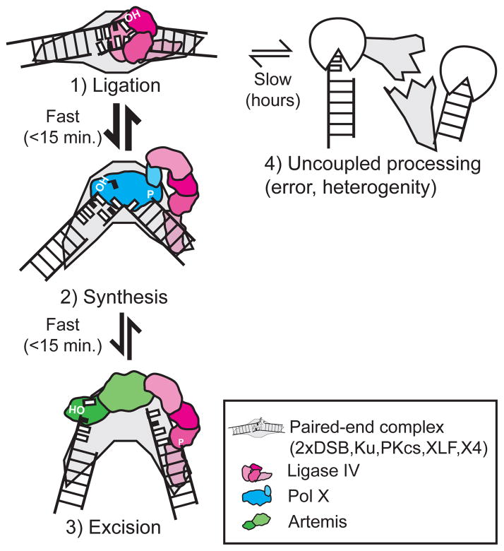
Nonhomologous end joining (NHEJ) can effectively resolve chromosome breaks despite diverse end structures; however, it is unclear how the steps employed for resolution are determined. We sought to address this question by analysing cellular NHEJ of ends with systematically mispaired and damaged termini. We show NHEJ is uniquely proficient at bypassing subtle terminal mispairs and radiomimetic damage by direct ligation. Nevertheless, bypass ability varies widely, with increases in mispair severity gradually reducing bypass products from 85 to 6%. End-processing by nucleases and polymerases is increased to compensate, although paths with the fewest number of steps to generate a substrate suitable for ligation are favoured. Thus, both the frequency and nature of end processing are tailored to meet the needs of the ligation step. We propose a model where the ligase organizes all steps during NHEJ within the stable paired-end complex to limit end processing and associated errors.


Conflict of interest statement

Competing Financial Interests

The authors declare no competing financial interests.

Figures

Figure 1
Figure 1. Design of substrates
a) Substrate TGCG3′ is shown; this and related substrates all possess symmetric head and tail end structures that can be aligned to allow for resolution by i) direct low-fidelity ligation of a terminal mispair, ii) gap fill-in synthesis and ligation, iii) excision of the terminal mispair, gap fill-in synthesis, and ligation (edit), or iv) other deletions. Nucleotides added during resolution are bolded. b) Left panel; TGCG3′ overhang substrate (generates G/T mispair; S) was incubated with Ku, XLF, XRCC4-LIG4 complex, and with and without Pol λ (left panel) to generate concatemer ligation products (P). Right panel; junctions were characterized by amplification and mock digested or digested with restriction enzymes diagnostic for direct ligation (Dir.) or synthesis and ligation (Syn.) (Supplementary Fig. 1a). c) A substrate with AGCG3′ overhangs (generates G/A mispair) analyzed as in panel b).
Figure 2
Figure 2. Cellular assay for NHEJ of systematically varied end structures
a) Description of cellular assay. b) Joining efficiencies for each substrate, comparing wild type HCT 116 cells to its LIG4 deficient variant (LIG4−/−). Error bars denote the standard deviation for 12 (5′GATC, 5′GCGT) or 6 (all others) independent electroporations. c) Substrates possessed end structure varied as shown. Product structures were defined as in Fig. 1a, and the proportion (%) of each determined by sequencing of junctions from wild type cells, averaged from two libraries, each library from a pool of 3 electroporations (see also methods). Error bars represent the range of results from the two libraries. d), e) The change in proportion of each resolution path due to differing terminal mispairs was calculated by subtracting mean proportions for each category of product, first (d)TGCG3′ (3′ G:T) from AGCG3′ (3′ G:A), then (e) 5′GCGT (5′G:T) from 5′GCGA (5′ G:A).
Figure 3
Figure 3. Characterization of junctions with deletions
a) Deletions were categorized according to whether deleted sequence was entirely limited to the single stranded overhangs (ssDNA deletion), then further categorized as whether the junction equaled the “edit” product described in Fig 1a, iii, vs. all other ssDNA deletions. Similarly, junctions where deleted sequence extended into flanking double stranded DNA (dsDNA deletion) then classed as occurring at a flanking sequence identity (microhomology mediated) or not (other dsDNA) b) Proportions of junctions with deletion (Fig. 1a, iii and iv) from Fig. 2c results were further categorized as in panel a). Error bars represent the range of results from the two libraries. c) The area of each slice is representative of the proportion of a different junction sequence with ssDNA deletion, as a fraction of the total sequences with ssDNA deletion (Fig. 3a, Supplementary Table 3). The proportion of edits (Fig. 1A, iii) is distinguished from deletions not guided by overhang sequence complementarity.
Figure 4
Figure 4. Changes in resolution path over time in the cell
a), and b) The change in proportion (%) of resolution path, comparing product recovered 5 hrs vs 15 minutes in cells, for 5′GCGT (a) and AGCG3′ (b). c) The frequency of different junctions, distinguishing accurate (filled section), microhomology directed deletions (diagonal bars), and all other deletions (open sections) for 5′GCGT vs AGCG3′ substrates, after 15 minutes versus 5 hours in cells.
Figure 5
Figure 5. NHEJ of systematically varied end structures in melanocytes
a) Selected substrates were introduced into melanocytes (NHM) and characterized as described in Fig. 2c, except results are from three independent libraries (9 electroporations). b), c) The change in proportion of each resolution path comparing NHM and HCT116 cells for substrates TGCG3′ (b) and 5′GCGT (c).
Figure 6
Figure 6. NHEJ of ends with a terminal 8 oxoguanine
a) Joining efficiencies for each substrate and cell line, all compared to joining of undamaged 5′GATC in wild type cells. Error bars represent the standard deviation from 6 independent electroporations. Joining in LIG4 deficient cells was significantly different for 5GOATC compared to the other two substrates (*, p<.05; ****, p<.0001; one way ANOVA comparing results of 6 independent electroporations for each group, with p values adjusted to account for multiple comparisons by the Bonferoni method). b) The proportion of junctions joined by noted resolution paths in HCT 116 cells after 15 minutes in cells, averaged from two libraries, each library from a pool of 3 electroporations. Error bars note the range. c), d) The change in proportion of each resolution path comparing 5 hrs vs 15 minutes, for HCT 116 cells (c) and melanocytes (d). e) Pathways for replacing GO with undamaged G.
Figure 7
Figure 7. Model for organization of enzymatic steps during NHEJ
A model for the order of steps and the configurations of ends and enzymes during repair by Nonhomologous end joining.

References

    1. O’Driscoll M, Jeggo PA. The role of double-strand break repair —insights from human genetics. Nat Rev Genet. 2006;7:45–54. - PubMed
    1. Lieber MR. The Mechanism of Double-Strand DNA Break Repair by the Nonhomologous DNA End-Joining Pathway. Annu Rev Biochem. 2010;79:181–211. - PMC - PubMed
    1. Dobbs TA, Palmer P, Maniou Z, Lomax ME, O’Neill P. Interplay of two major repair pathways in the processing of complex double-strand DNA breaks. DNA Repair (Amst ) 2008;7:1372–1383. - PubMed
    1. Datta K, Purkayastha S, Neumann RD, Pastwa E, Winters TA. Base damage immediately upstream from double-strand break ends is a more severe impediment to nonhomologous end joining than blocked 3′-termini. Radiation research. 2011;175:97–112. - PMC - PubMed
    1. Zhou RZ, et al. Tolerance for 8-oxoguanine but not thymine glycol in alignment-based gap filling of partially complementary double-strand break ends by DNA polymerase lambda in human nuclear extracts. Nucleic Acids Res. 2008;36:2895–2905. - PMC - PubMed

Publication types

LinkOut - more resources