. 2014 Sep 1;25(17):2604-19.
doi: 10.1091/mbc.E14-02-0712. Epub 2014 Jul 2.

Ena/VASP regulates mDia2-initiated filopodial length, dynamics, and function

Affiliations

Ena/VASP regulates mDia2-initiated filopodial length, dynamics, and function

Melanie Barzik et al. Mol Biol Cell. .

Abstract

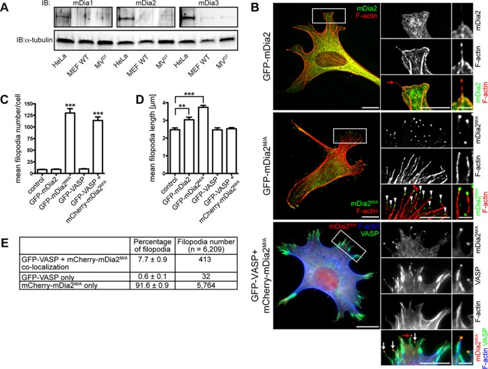
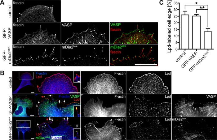
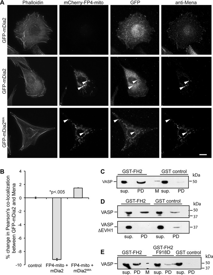
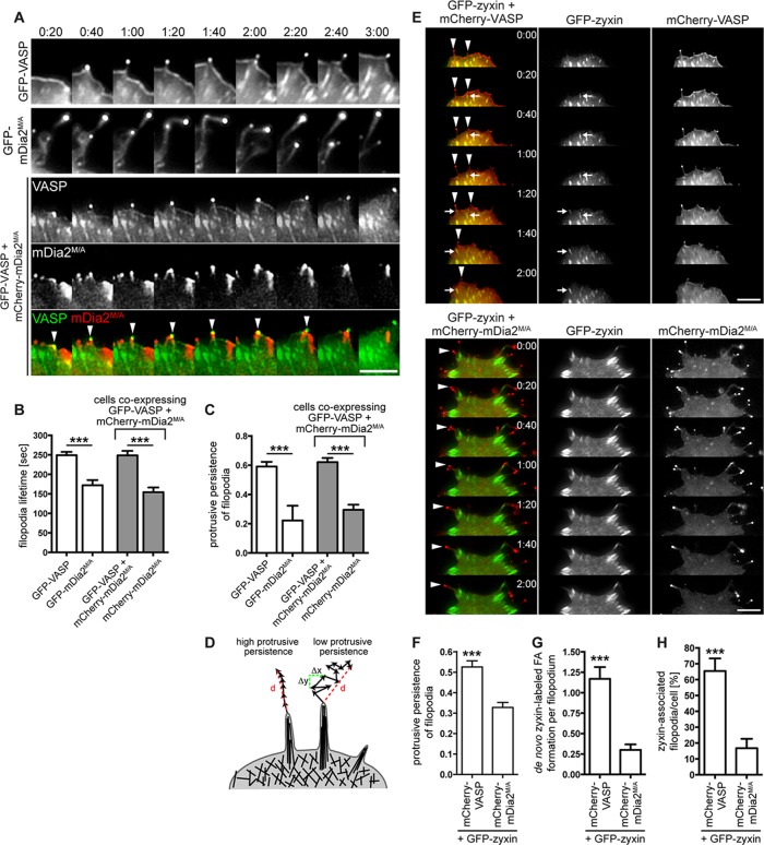
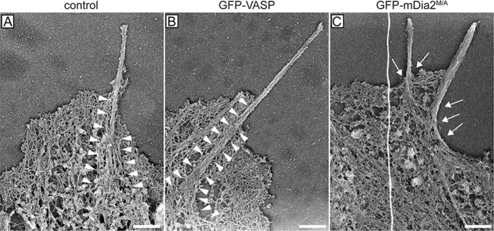
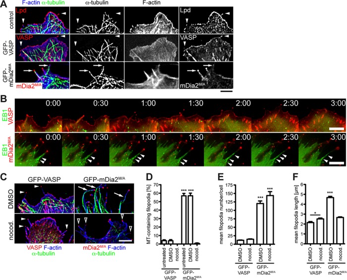
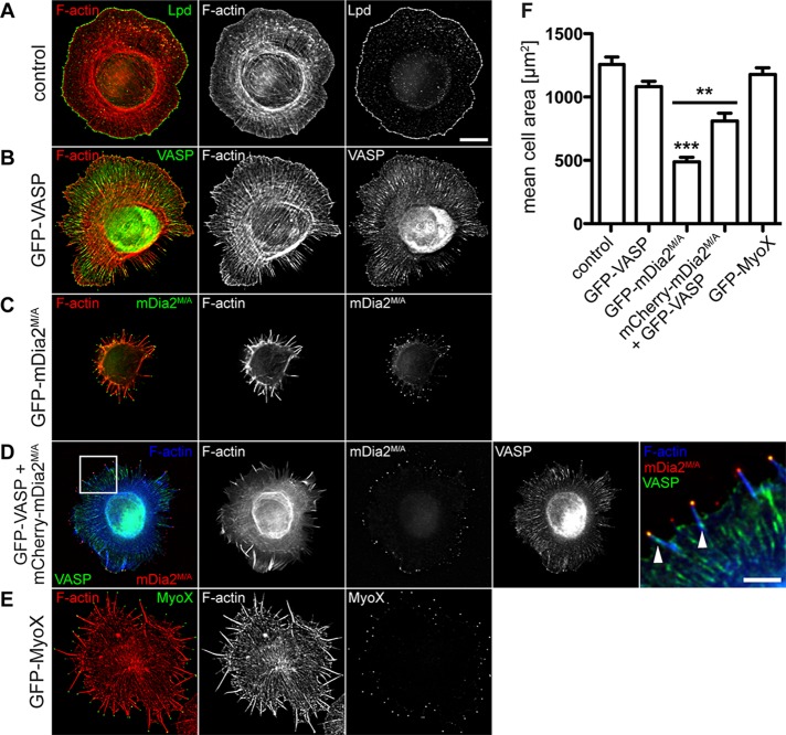
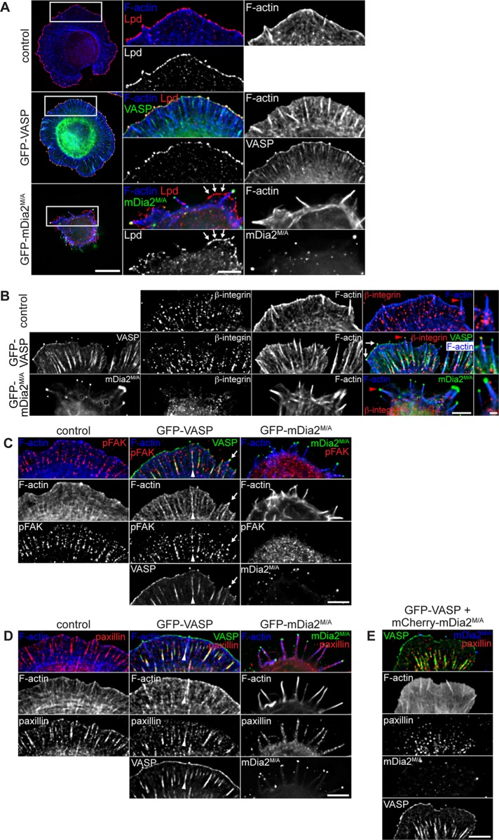
Filopodia are long plasma membrane extensions involved in the formation of adhesive, contractile, and protrusive actin-based structures in spreading and migrating cells. Whether filopodia formed by different molecular mechanisms equally support these cellular functions is unresolved. We used Enabled/vasodilator-stimulated phosphoprotein (Ena/VASP)-deficient MV(D7) fibroblasts, which are also devoid of endogenous mDia2, as a model system to investigate how these different actin regulatory proteins affect filopodia morphology and dynamics independently of one another. Filopodia initiated by either Ena/VASP or mDia2 contained similar molecular inventory but differed significantly in parameters such as number, length, F-actin organization, lifetime, and protrusive persistence. Moreover, in the absence of Ena/VASP, filopodia generated by mDia2 did not support initiation of integrin-dependent signaling cascades required for adhesion and subsequent lamellipodial extension, thereby causing a defect in early cell spreading. Coexpression of VASP with constitutively active mDia2(M/A) rescued these early adhesion defects. We conclude that Ena/VASP and mDia2 support the formation of filopodia with significantly distinct properties and that Ena/VASP regulates mDia2-initiated filopodial morphology, dynamics, and function.


Figures

FIGURE 1:
FIGURE 1:
Active mDia2 induces abundant long filopodia in the absence of VASP. (A) MVD7 cells lack endogenous mDia1 and mDia2. Cell extract from MVD7 cells was analyzed by SDS–PAGE and immunoblot using antibodies specific for mDia1, mDia2, or mDia3. Controls: extracts of wild-type MEFs and HeLa cells (which endogenously express mDia proteins); loading control: α-tubulin. (B) Effect of VASP and mDia2 on filopodia formation. MVD7 cells or MVD7 cells stably expressing GFP-VASP were transiently transfected with the mDia2 constructs as indicated and were chemically fixed after 24 h and probed with phalloidin to label F-actin. As expected, GFP-VASP localization to focal adhesions, lamellipodia, and filopodia tips was observed. Boxed regions represent magnified area; red arrows/arrowheads indicate filopodia magnified on the right. (C–E) Quantification of B showing mean filopodia number (C), mean length of filopodia (D), and percentage of VASP and mDia2M/A colocalization at filopodia tips in cells coexpressing GFP-VASP and mCherry-mDia2M/A (E). Data in C–E represent at least two repetitions. ***p ≤ 0.001, **p ≤ 0.01; n = 33–59 cells. Scale bars, 15 μm; magnified regions, 5 μm; single filopodia, 1 μm.
FIGURE 2:
FIGURE 2:
MVD7 cells expressing mDia2M/A have smaller lamellipodia. (A) MVD7 cells expressing mDia2M/A contain the normal filopodia inventory. MVD7 cells producing the indicated proteins were chemically fixed and stained for the actin cross-linking protein fascin. (B) Lpd antibody labels filopodia tips (arrows) and the actively protruding leading edge of lamellipodia. Right, magnification of boxed regions. Red arrows indicate filopodia magnified in insets. (C) Quantification of Lpd-labeled cell edge from experiments shown in B. Cells were transiently transfected, fixed, and stained with Lpd antibody. Data in C represent at least two repetitions. *p ≤ 0.05, **p ≤ 0.01; n = 10–13 cells. Scale bars, 15 μm; magnified images, 5 μm; single-filopodium insets, 1 μm.
FIGURE 3:
FIGURE 3:
VASP directly interacts with mDia2. (A) Mitochondria-targeting assay. Transiently transfected NIH3T3 cells producing the indicated GFP- or mCherry-tagged proteins were chemically fixed and probed with a marker for F-actin (phalloidin) or antibody directed against Mena. Arrowheads indicate mitochondria labeled with mCherry-FP4-mito. Scale bar, 15 μm. (B) Quantification of A showing percentage change in Pearson's colocalization coefficient between Mena and GFP-mDia2. (C–E) Pull-down studies analyzing complex formation between mDia2 and VASP. Bead-immobilized GST-FH2 domain was incubated with purified VASP or VASPΔEVH1, and protein retained on beads was detected by SDS–PAGE and immunoblot. sup., supernatant; PD, pull down; M, molecular weight marker. (C) VASP interacts with the mDia2 FH2 domain. (D) VASP binds mDia2 through its EVH1 domain. (E) The FPPP motif in mDia2 FH2 mediates interaction with VASP. All data are from at least three independent experiments.
FIGURE 4:
FIGURE 4:
VASP is essential for persistent protrusion of mDia2-generated filopodia. (A) VASP restores protrusive behavior of mDia2M/A-induced filopodia. MVD7 cells were transfected as indicated and plated on fibronectin-coated glass bottom dishes for 24 h before live-cell imaging. Montages of single frames from 3:00 min of live-cell microscopy time series. (B, C) Quantification of protrusive persistence (B) and lifetime (C) of filopodia from experiments shown in A. Filopodia tips in cells expressing GFP-VASP, GFP-mDia2M/A, or GFP-VASP plus mCherry-mDia2M/A were tracked in individual frames of 5-min movies, and so were filopodia that were in the same coexpressing cells but that contained only mCherry-mDia2M/A. Data represent at least two repetitions. n = 7–16 cells. (D) Distinct filopodia dynamics results in variable protrusive persistence. Protrusive persistence of filopodia was calculated by measuring the total distance (d) traveled by the filopodium tip divided by the sum of the change in directionality (Δx, Δy) of the filopodium tip between individual frames (see Materials and Methods). (E–H) VASP- but not mDia2M/A-containing filopodia (arrowheads) induce de novo assembly of GFP-zyxin–labeled focal adhesions at their base or in the shaft while actively protruding (arrows). MVD7 cells were transfected as indicated and plated on fibronectin-coated glass-bottom dishes for 24 h before live-cell imaging. (E) Montages of single frames from 2 min of live-cell microscopy time series. (F) GFP-zyxin expression does not change filopodia dynamics. (G) Quantification of de novo focal adhesion formation in protruding filopodia from experiments shown in E. (H) Percentage of zyxin-associated filopodia/cell. Data in F–H represent at least two repetitions. N = 8–11 cells, n = 41–60 filopodia in F and G; n = 58 (VASP) and 1426 (mDia2M/A) filopodia in H. ***p < 0.001. Scale bars, 5 μm.
FIGURE 5:
FIGURE 5:
Actin bundles in filopodia initiated by mDia2M/A are not anchored in the lamellipodium actin network. Representative platinum-replica electron micrographs of actin cytoskeletons from MVD7 cells. Filopodium F-actin bundles from control cells (A) and cells complemented with GFP-VASP (B) are deeply embedded in the actin network of the lamellipodium (arrowheads) and splay apart at the filopodium root. In contrast, filopodia actin bundles of cells producing GFP-mDia2M/A (C) display poor connection to the underlying cortical cytoskeleton and do not have splayed roots (arrows), indicating that they may have formed via a different mechanism. Electron micrographs are representative images from at least three independent experiments. Scale bars, 0.5 μm.
FIGURE 6:
FIGURE 6:
Microtubule targeting of mDia2M/A-initiated filopodia. (A) mDia2M/A-assembled filopodia contain microtubules. MVD7 cells were transiently transfected, chemically fixed, and probed for microtubules (α-tubulin), filopodia (Lpd), and F-actin (phalloidin). Arrows indicate microtubules extending into tips of mDia2M/A-assembled filopodia. Arrowheads show that microtubules do not extend into filopodia of control or GFP-VASP–expressing cells. (B) Actively polymerizing microtubules perpetually protrude into mDia2M/A-induced filopodia. Montages of single frames from 3-min time series of cells coexpressing mCherry-EB1 and either GFP-VASP (top) or GFP-mDia2M/A (bottom). Arrowheads indicate EB1-labeled microtubules actively entering mDia2M/A-labeled filopodia. (C) Filopodia in GFP-VASP–expressing (left) or GFP-mDia2M/A–expressing (right) cells are unaffected by microtubule depolymerization. Transfected cells were exposed to 100 ng/ml nocodazole for 15 h (DMSO control, top; nocodazole, bottom) and probed for α-tubulin and F-actin (phalloidin). Nocodazole does not affect filopodia formation in GFP-VASP–expressing cells (filled arrowheads). DMSO treatment does not prevent microtubules from entering GFP-mDia2M/A–labeled filopodia (arrows). Depolymerization of microtubules does not inhibit filopodia formation in cells expressing GFP-mDia2M/A but reduces filopodial length (open arrowheads). (D–F) Quantification of the experiments shown in A and C. (D) mDia2M/A-labeled filopodia do not contain microtubules in nocodazole-treated cells. (E) Microtubules are not required for GFP-mDia2M/A–driven filopodia formation. (F) Microtubule depolymerization dramatically reduces filopodia length in cells expressing GFP-mDia2M/A, whereas filopodial length in GFP-VASP–expressing cells is slightly increased. Data in D–F represent at least two repetitions. *p < 0.05; ***p < 0.001. DMSO, vehicle control; nocod., nocodazole. n = 39–55 cells. Scale bars, 5 μm.
FIGURE 7:
FIGURE 7:
Filopodia initiated by mDia2M/A do not support early cell spreading. (A–E) Cell spreading assay. At 24 h after transfection, MVD7 control cells (A) or MVD7 cells producing (B) GFP-VASP, (C) GFP-mDia2M/A, (D) GFP-VASP + mCherry-mDia2M/A, or (E) GFP-MyoX were allowed to spread on laminin-coated coverslips for 30 min and fixed, and F-actin was labeled with phalloidin. Control cells were also probed with antibody directed against Lpd. Box in D shows localization of magnified region on the right, where small focal adhesions labeled by GFP-VASP at the base of filopodia are indicated by arrowheads. (F) Quantification of cell area from cell spreading assays as depicted in A–E. Data represent at least two independent experiments. ***p < 0.001; **p < 0.01. n = 43-45 cells. Scale bar, 15 μm; magnified image, 5 μm.
FIGURE 8:
FIGURE 8:
mDia2M/A-initiated filopodia lack protrusive and adhesive structures. At 24 h posttransfection, MVD7 cells were allowed to spread on laminin for 30 min, fixed, and immunostained for (A) Lpd, (B) activated β1 integrin subunit, (C) activated FAK (pFAK), or (D) paxillin. (A) Lamellipodium formation in spreading GFP-mDia2M/A–expressing cells is dramatically reduced (arrows). Right, magnification of boxed regions. (B) β1 integrin is not activated in tips of mDia2M/A-containing filopodia. Activated β1 integrin colocalizes with GFP-VASP to filopodia tips (arrows). Red arrowheads indicate individual filopodia magnified on the right. (C) Activated FAK is not recruited to filopodia tips or focal adhesions in GFP-mDia2M/A–expressing cells. In contrast, pFAK and GFP-VASP colocalize at filopodia tips (arrows) and in focal adhesions at the base of VASP-labeled filopodia (arrowheads). (D) Paxillin-containing focal adhesions are absent in spreading GFP-mDia2M/A–expressing MVD7 cells but are formed at the base of VASP-labeled filopodia (arrowheads). (E) Formation of elongated paxillin-containing focal adhesions in spreading mDia2M/A-expressing cells is rescued in the presence of VASP. All images are from at least two independent experiments. Scale bars, 10 μm; magnified images, 5 μm; magnified images of single filopodia in B, 1 μm.

Similar articles

Cited by

References

    1. Alberts AS. Identification of a carboxyl-terminal diaphanous-related formin homology protein autoregulatory domain. J Biol Chem. 2001;276:2824–2830. - PubMed
    1. Alexandrova AY, Arnold K, Schaub S, Vasiliev JM, Meister JJ, Bershadsky AD, Verkhovsky AB. Comparative dynamics of retrograde actin flow and focal adhesions: formation of nascent adhesions triggers transition from fast to slow flow. PloS One. 2008;3:e3234. - PMC - PubMed
    1. Applewhite DA, Barzik M, Kojima S, Svitkina TM, Gertler FB, Borisy GG. Ena/VASP proteins have an anti-capping independent function in filopodia formation. Mol Biol Cell. 2007;18:2579–2591. - PMC - PubMed
    1. Arias-Salgado EG, Lizano S, Sarkar S, Brugge JS, Ginsberg MH, Shattil SJ. Src kinase activation by direct interaction with the integrin beta cytoplasmic domain. Proc Natl Acad Sci USA. 2003;100:13298–13302. - PMC - PubMed
    1. Aspenström P. Formin-binding proteins: modulators of formin-dependent actin polymerization. Biochim Biophys Acta. 2010;1803:174–182. - PubMed

Publication types

MeSH terms

LinkOut - more resources