. 2014 Oct;32(10):2702-13.
doi: 10.1002/stem.1784.

Leptin signaling is required for augmented therapeutic properties of mesenchymal stem cells conferred by hypoxia preconditioning

Affiliations

Leptin signaling is required for augmented therapeutic properties of mesenchymal stem cells conferred by hypoxia preconditioning

Xinyang Hu et al. Stem Cells. 2014 Oct.

Erratum in

  • Stem Cells. 2014 Dec;32(12):3287

Abstract

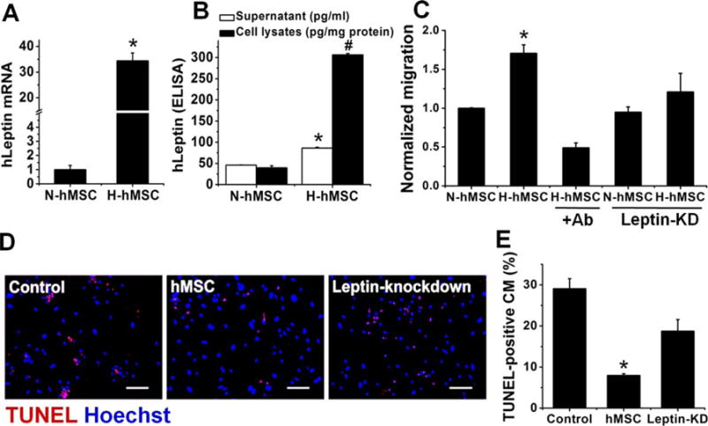
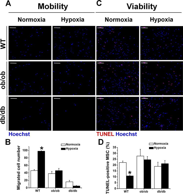
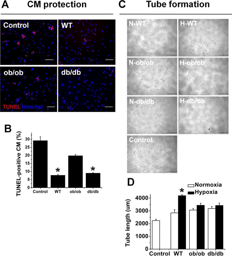
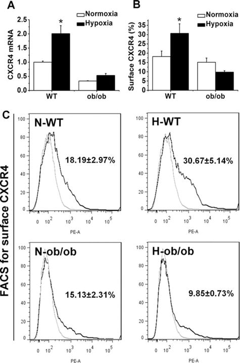
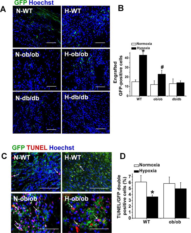
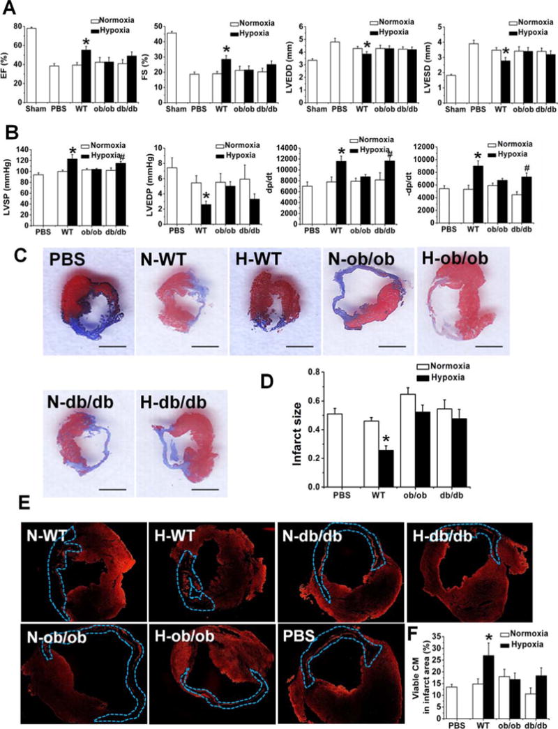
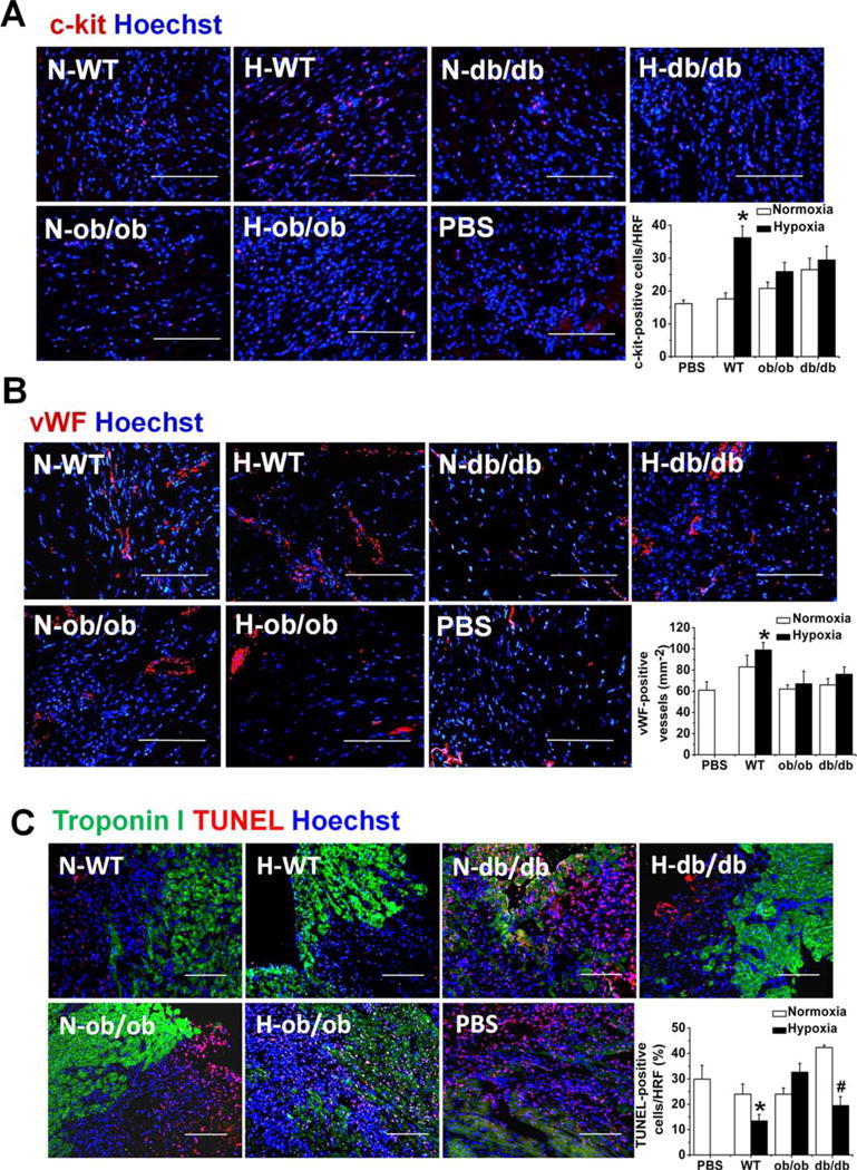
Hypoxia preconditioning enhances the therapeutic effect of mesenchymal stem cells (MSCs). However, the mechanism underlying hypoxia-induced augmentation of the protective effect of MSCs on myocardial infarction (MI) is poorly understood. We show that hypoxia-enhanced survival, mobility, and protection of cocultured cardiomyocytes were paralleled by increased expression of leptin and cell surface receptor CXCR4. The enhanced activities were abolished by either knockdown of leptin with a selective shRNA or by genetic deficiency of leptin or its receptor in MSCs derived, respectively, from ob/ob or db/db mice. To characterize the role of leptin in the regulation of MSC functions by hypoxia and its possible contribution to enhanced therapeutic efficacy, cell therapy using MSCs derived from wild-type, ob/ob, or db/db mice was implemented in mouse models of acute MI. Augmented protection by hypoxia pretreatment was only seen with MSCs from wild-type mice. Parameters that were differentially affected by hypoxia pretreatment included MSC engraftment, c-Kit(+) cell recruitment to the infarct, vascular density, infarct size, and long-term contractile function. These data show that leptin signaling is an early and essential step for the enhanced survival, chemotaxis, and therapeutic properties of MSCs conferred by preculture under hypoxia. Leptin may play a physiological role in priming MSCs resident in the bone marrow endosteum for optimal response to systemic signaling molecules and subsequent tissue repair.

Keywords: CXCR4; Hypoxic preconditioning; Leptin; Mesenchymal stem cells; Myocardial infarction.


Conflict of interest statement

of Potential Conflicts of Interest The authors indicate no potential conflicts of interest.

Figures

Figure 1
Figure 1
Leptin production and cellular properties of hMSCs. (A): Leptin mRNA measured by quantitative real-time PCR of total RNA isolated from human MSCs cultured at normoxia (N-hMSC) and hypoxia (H-hMSC) condition for 24 hours. mRNA value of H-hMSC was normalized to N-hMSC (n=3, *, p<.05). (B): Leptin protein from human MSC was measured by ELISA. Cell lysates and culture supernatant were collected from 8×10⁵ cells hMSCs after cultured with 3 ml medium for 24 hours at either normoxia and hypoxia condition (n=3, * #, p<.05 vs. N-hMSC). (C): Comparison of migration of hMSC and leptin-knockdown hMSC after normoxia or hypoxia pretreatment. +Ab is H-hMSC cultured with leptin neutralized antibody. Leptin-KD is that hMSCs were transduced with lentiviral vector carrying shRNA specific for leptin (Leptin-knockdown) (n=3, *, p<.05 vs. others). (D): Apoptosis of CMs from neonatal mouse. CMs were cocultured with no cell (Control), or hMSC without modification (hMSC) or hMSC with Leptin-knockdown under condition of simulated ischemia for 48 hours. CMs were subjected TUNEL staining (red) for apoptotic cells and Hoechst 33258 staining for nuclei (blue). Bars represent 100 μm. (E): Quantification of apoptotic cardiomyocytes in D (n=3, *, p<.05 vs. Control and Leptin-knockdown hMSC). Data are represented as mean ± SE. Abbreviations: CM, cardiomyocyte; ELISA, enzyme-linked immunosorbent assay; hMSC, human mesenchymal stem cell.
Figure 2
Figure 2
Comparison of mobility and viability among mouse MSCWT, MSCob/ob, and MSCob/ob in vitro. (A): Transwell assay of migration of MSCs after normoxia or hypoxia pretreatment. Migrated cells were stained with Hoechst and visualized under fluorescent microscopy. (B): Quantification of migrated cells (n=3, *, p<.05 vs. others). (C): TUNEL staining detecting apoptotic MSCs after normoxia or hypoxia pretreatment for 24 hours and then cultured in a simulated ischemia condition. Total MSCs were identified by Hoechst 33258 staining for nuclei (blue), and apoptotic cells were TUNEL positive (red). (D): Quantification of apoptotic cells (n=3, *, p<.05 vs. others). Bars in A and C represent 100 μm. Abbreviations: MSC, mesenchymal stem cell; WT, wild type.
Figure 3
Figure 3
Comparison of the paracrine effects of different mesenchymal stem cells (MSCs) in vitro. (A): CM protection was determined by measuring apoptosis of CMs that were cocultured with MSCs. CMs from neonatal mouse were cocultured with no cell (Control), or MSCWT (WT) or MSCob/ob (ob/ob) or MSCdb/db (db/db) under condition of simulated ischemia for 48 hours. CMs were subjected TUNEL staining (red) for apoptotic cells and Hoechst 33258 staining for nuclei (blue). (B): Quantification of apoptotic CMs (n=3, *, p<.05 vs. Control and ob/ob). (C): Proangiogenesis effect of MSC was determined by examining tube formation of HUVEC cultured with conditioned medium from no cell (Control) and MSCs. MSCWT (WT), MSCob/ob (ob/ob), or MSCdb/db (db/db) were cultured at normoxia or hypoxia condition for 24 hours before their conditioned media were collected to be cultured with HUVEC in Matrigel for 4 hours. (D): Quantification of tube length in C (n=3, *, p<.05 vs. Control, N-WT, H-ob/ob, and H-db/db). Bars in A and C represent 100 μm. Abbreviations: CM, cardiomyocyte; WT, wild type.
Figure 4
Figure 4
CXCR4 expression in MSCWT and MSCob/ob. (A): CXCR4 mRNA measured by quantitative real-time PCR of total RNA isolated from MSCWT and MSCob/ob after cultured at normoxia and hypoxia for 24 hours. mRNA values were normalized to N-MSCWT (n=3, *, p<.05 vs. N-WT and H-ob/ob). (B): Quantification of surface CXCR4 expression on MSCWT and MSCob/ob (n=3, *, p<.05 vs. N-WT and H-ob/ob) measured by flow cytometry shown in C. (C): FACS analysis of CXCR4 surface expression on mesenchymal stem cells (MSCs) after cultured at normoxia and hypoxia for 24 hours. Gray lines represent controls with isotype Ab, black lines represent data from PE-conjugated Ab. Abbreviations: FACS, fluorescence-activated cell sorting; WT, wild type.
Figure 5
Figure 5
Mesenchymal stem cell (MSC) homing and apoptosis in vivo. (A): Comparison of homing potential among different mouse MSCs after normoxia or hypoxia pretreatment. MSCWT (WT) or MSCob/ob (ob/ob) or MSCdb/db (db/db) were transduced with a GFP-lentiviral vector and then injected via tail vein into mice with myocardial infarction (MI) surgery. The hearts were harvested 2 days later, and sections were immunostained for GFP (green) and Hoechst 33258 for nuclei (blue). (B): Quantification of engrafted GFP+ cells (n=3 for N-WT, H-WT, N-ob, H-ob, N-db, 4 H-db, *, p<.05 vs. N-WT, H-ob/ob and H-db/db, #, p<.05 vs. N-ob/ob). (C): The retention and survival of normoxic or hypoxic MSCWT/MSCob/ob in border zone of LV infarct at 3 days after MI. TUNEL staining for detecting apoptosis (red) of MSC (GFP-positive, green fluorescence) and Hoechst (blue) for nuclear staining. (D): Quantification of apoptotic GFP cells (GFP/TUNEL double-positive) at 3 days after MI (n=4, *, p<.05 vs. N-WT and H-ob/ob). The percentage of TUNEL/GFP double positive cells was calculated by dividing the number of TUNEL/GFP double positive cells with the total GFP-positive cell number. Bars in A and C represent 100 μm. Abbreviations: GFP, green fluorescent protein; WT, wild type.
Figure 6
Figure 6
Protection of cardiac function after transplantation of mesenchymal stem cells (MSCs) into the infarcted heart. PBS, or MSCWT, MSCob/ob, or MSCdb/db preconditioned with normoxia or hypoxia were injected into the peri-infarct zones after myocardial infarction (MI). Four weeks after MI. (A): Echocardiographic examination was performed, and EF, FS, LVEDD, LVESD were obtained to evaluate the cardiac function in each groups; n=7 for Sham, 8 PBS, 6 N-WT, 8 H-WT, 12 N-ob, 15 H-ob, 13 N-db, 14 H-db. *, p<.05 versus others. (B): Hemodynamic analyses at 4 weeks post-MI. Left ventricular pressure was recorded and hemodynamic parameter including LVSP, LVEDP, and both maximum positive and negative rate of changes in pressure (±dp/dtmax) were also obtained, demonstrating improved cardiac function after cell transplantation. n=13 for PBS, 7 N-WT, 7 H-WT, 9 N-ob, 17 H-ob. *, p<.05 versus PBS, N-WT, and ob; #, p<.05 versus PBS, N-db. (C): Representative Masson’s trichrome staining of heart tissue to show the infarct area (blue). (D): Quantification of infarct size. n=6 for PBS, 5 N-WT, 5 H-WT, 6 N-ob, 5 H-ob, 6 N-db, 5 H-db. *, p<.05 versus others. (E): Troponin I-immunostaining of the recovered heart sections for viable cardiomyocytes. Dot lines show the infarct area in which the viable CMs were quantified in F. (F): Quantification of viable CMs. n=4 for PBS, 4 N-WT, 4 H-WT, 5 N-ob, 4 H-ob, 5 N-db, 3 H-db. *, p<.05 versus others. Abbreviations: CM, cardiomyocyte; EF, ejection fraction; FS, fraction shortening; LVEDD, left ventricular end-diastolic diameter; LVESD, left ventricular end-systolic diameter; LVSP, left ventricular systolic pressure; LVEDP, left ventricular end diastolic pressure; WT, wild type.
Figure 7
Figure 7
Effect of mesenchymal stem cells on c-kit-positive cardiac progenitor cells, endothelial cells, and cardiac cells. Heart tissues (LV) recovered 4 weeks after MI and cell therapy were subjected for immunofluorescence staining. (A): Recruitment of cardiac progenitor cells (c-kit-positive) in border zone of infarct LV at 3 days post-MI. n=6 for PBS, 9 N-WT, 8 H-WT, 6 N-ob, 7 H-ob, 4 N-db, 6 H-db. (B): Capillary density (vWF-positive) in border zone of LV infarct at 28 days post-MI. ECs were identified by staining for vWF. n=4. (C): TUNEL staining for apoptotic cardiomyocytes at the board zone of infarct LV at 3 days after MI. n=5 for PBS, 9 N-WT, 6 H-WT, 5 N-ob, 5 H-ob, 3 N-db, 7 H-db. The bar graphs are the quantification of the results from A to C. *, p<.05 versus others. #, p<.05 versus normoxic-db/db. Bars in the pictures represent 100 μm. Abbreviations: HRF, high resolution field; WT, wild type.

Similar articles

Cited by

References

    1. van Ramshorst J, Rodrigo SF, Schalij MJ, et al. Bone marrow cell injection for chronic myocardial ischemia: The past and the future. J Cardiovasc Transl Res. 2011;4:182–191. - PMC - PubMed
    1. Ptaszek LM, Mansour M, Ruskin JN, et al. Towards regenerative therapy for cardiac disease. Lancet. 2012;379:933–942. - PubMed
    1. Donndorf P, Strauer B-E, Steinhoff G. Update on cardiac stem cell therapy in heart failure. Curr Opin Cardiol. 2012;27:154–160. - PubMed
    1. Hung SC, Pochampally RR, Hsu SC, et al. Short-term exposure of multipotent stromal cells to low oxygen increases their expression of CX3CR1 and CXCR4 and their engraftment in vivo. PLoS One. 2007;2:e416. - PMC - PubMed
    1. Liu H, Liu S, Li Y, et al. The role of SDF-1-CXCR4/CXCR7 axis in the therapeutic effects of hypoxia-preconditioned mesenchymal stem cells for renal ischemia/reperfusion injury. PLoS One. 2012;7:e34608. - PMC - PubMed

Publication types