. 2014 Sep 2;112(3):558-65.
doi: 10.1160/TH13-12-1013. Epub 2014 Jul 3.

Thrombin-induced platelet activation via PAR4: pivotal role for exosite II

Affiliations

Thrombin-induced platelet activation via PAR4: pivotal role for exosite II

N Boknäs et al. Thromb Haemost. .

Abstract

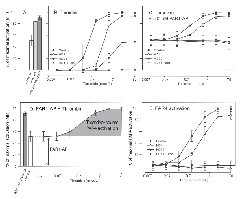
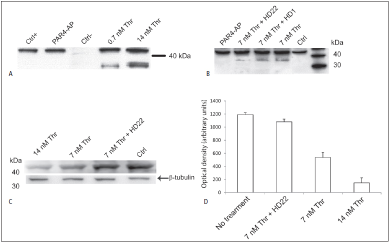
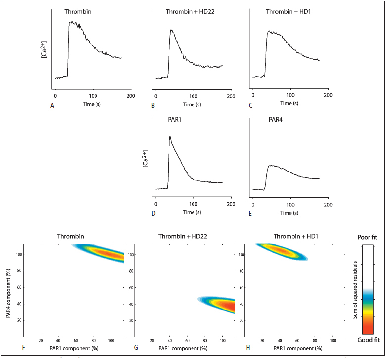
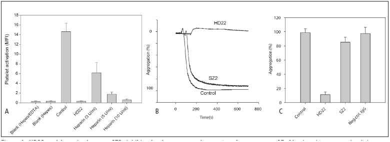
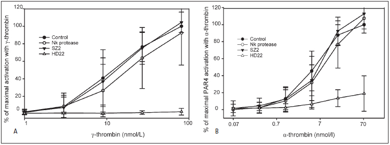
Thrombin-induced platelet activation via PAR1 and PAR4 is an important event in haemostasis. Although the underlying mechanisms responsible for ensuring efficient PAR1 activation by thrombin have been extensively studied, the potential involvement of recognitions sites outside the active site of the protease in thrombin-induced PAR4 activation is largely unknown. In this study, we developed a new assay to assess the importance of exosite I and II for PAR4 activation with α - and γ-thrombin. Surprisingly, we found that exosite II is critical for activation of PAR4. We also show that this dependency on exosite II likely represents a new mechanism, as it is unaffected by blockage of the previously known interaction between thrombin and glycoprotein Ibα.


Conflict of interest statement

Conflicts of interest None declared.

Figures

Figure 1: Activation of PAR4 by α-thrombin; critical dependence on exosite II
. A) Platelet-rich plasma mixed with FITC-conjugated PAC-1 antibody was incubated with 100 μM PAR1 -AP (first column), 500 μM PAR4-AP (second column) and 100 μM PAR1-AP plus 500 μM PAR4-AP (third column) for 16 min, after which the mean fluorescence intensity (MFI) was measured with flow cytometry. B) Platelets were incubated for 16 min with increasing concentrations of thrombin, either alone or preincubated with 1 μM HD1, 1 μM HD22 or 1 μM HD1 plus 1 μM HD22. C) The experiment in (B) was repeated in the presence of 100 μM PAR1-AP to study the contribution of PAR4. D) PAR4-mediated platelet activation was calculated from the results in (B-C) by subtracting the signal obtained with 100 μM PAR1-AP from the signal with thrombin in the presence of 100 μM PAR1-AP. The normalised PAR4-mediated activation calculated by this method is shown in (E). Results in (A-D) are expressed as a percentage of the maximal MFI obtained with the control sample for each donor (thrombin in the concentration range 0.007-70 nmol/l). In (E), results are expressed as a percentage of the maximal calculated PAR4 activation obtained with the control sample. Data represent mean ± SD, n≥3.
Figure 2: PAR4 cleavage occurs at low thrombin concentrations and is inhibited by HD22
. Immunoblotting with the antibody 5F4 was performed on lysates of washed platelets. A) Platelets were exposed to 500 μM PAR4-AP, 0.7 nM α-thrombin or 14 nM α-thrombin. B) The relative density of a lower band representing cleaved PAR4 receptors was assessed after treatment with 7 nM thrombin alone or pre-incubated with the aptamers HD1 and HD22. C) The density of the upper band representing uncleaved PAR4 receptors was assessed after incubation with 7 nM α-thrombin alone or preincubated with HD22. D) The average band densities ± SD for three independent blots in (C), are shown as measured by densitometry. The estimated protein loading was 10 μg in (A) and 20 μg in (B-C). The shown blots are one representative example out of three independent experiments.
Figure 3: Kinetic profiles of thrombin-triggered calcium transients reveal differential inhibition of PAR receptors with HD1 and HD22
. A-C) Changes in intracellular Ca 2+ upon thrombin stimulation (7 nM) alone or in the presence of 1 μM HD22 or HD1 were recorded. D-E) Ca 2+ profiles obtained with 100 μM PAR1-AP alone or 500 μM PAR4-AP alone are provided for comparison. F-H) A combination matrix of the PAR1 and PAR4 calcium response profiles with variation in amplitude (0–100 %) were fitted to the calcium signal induced by thrombin with or without HD1 or HD22, where best fit can be seen in red as the minimum sum of squared residuals. Data represent the average of three independent experiments conducted on platelets from different donors.
Figure 4: HD22 and heparin, but not SZ2, inhibit platelet aggregation induced by -thrombin
. A) Washed platelets incubated with PAC-1 antibody were exposed to 27 nM γ-thrombin alone or together with 1 nM HD22, or the indicated concentrations of heparin for 16 min, after which the mean fluorescence intensity (MFI) was measured with flow cytometry. B-C) Aggregation after exposure to 27 nM γ-thrombin, measured as light transmission, was recorded for untreated PRP, and PRP pre-incubated with 1 μM HD22, 20 μg/ml control mAb or 20 μg/ml SZ2. Data represent mean ± SD (n = 3–5) in (A) and (C), whereas (B) shows one representative original trace.
Figure 5: GpIbα does not support PAR4 activation with α- or γ-thrombin
. Washed platelets incubated with PAC-1 antibody were exposed to different concentrations of (A) α-thrombin or (B) γ-thrombin. B) Titrations were performed in the presence or absence of PAR1 -AP (100 μM). The PAR4 response shown in (B) was then calculated as described in Figure 1 . Titrations were performed without further treatment (control), in the presence of 20 μg/ml SZ2, after treatment with Nk protease for depletion of GpIbα, or after incubation with 1 μM of HD22. Results are expressed as a percentage of the maximal mean fluorescence intensity obtained in each thrombin titration series. Each experiment was repeated at least three times with blood from different donors. Data represent mean ± SD.

Similar articles

Cited by

References

    1. Kahn ML, Zheng Y-W, Huang W. A dual thrombin receptor system for platelet activation. Nature. 1998;394:690–694. - PubMed
    1. Jonnalagadda D, Izu LT, Whiteheart SW. Platelet secretion is kinetically heterogeneous in an agonist-responsive manner. Blood. 2012;120:5209–5216. - PMC - PubMed
    1. Shapiro MJ, Weiss EJ, Faruqi TR. Protease-activated receptors 1 and 4 are shut off with distinct kinetics after activation by thrombin. J Biol Chem. 2000;275:25216–25221. - PubMed
    1. De Candia E, Hall SW, Rutella S. Binding of Thrombin to Glycoprotein Ib Accelerates the Hydrolysis of Par-1 on Intact Platelets. J Biol Chem. 2001;276:4692–4698. - PubMed
    1. Gandhi PS, Chen Z, Di Cera E. Crystal Structure of Thrombin Bound to the Un-cleaved Extracellular Fragment of PAR1. J Biol Chem. 2010;285:15393–15398. - PMC - PubMed

Publication types

MeSH terms