. 2014 Dec 1;23(23):6246-59.
doi: 10.1093/hmg/ddu344. Epub 2014 Jul 2.

Tissue-specific insulator function at H19/Igf2 revealed by deletions at the imprinting control region

Affiliations

Tissue-specific insulator function at H19/Igf2 revealed by deletions at the imprinting control region

Folami Y Ideraabdullah et al. Hum Mol Genet. .

Abstract

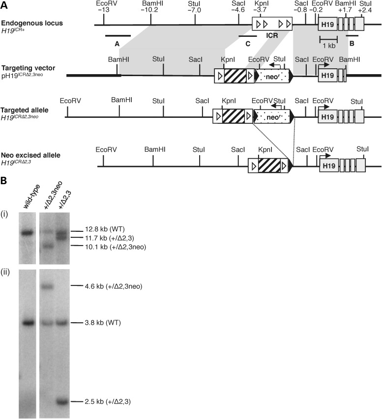
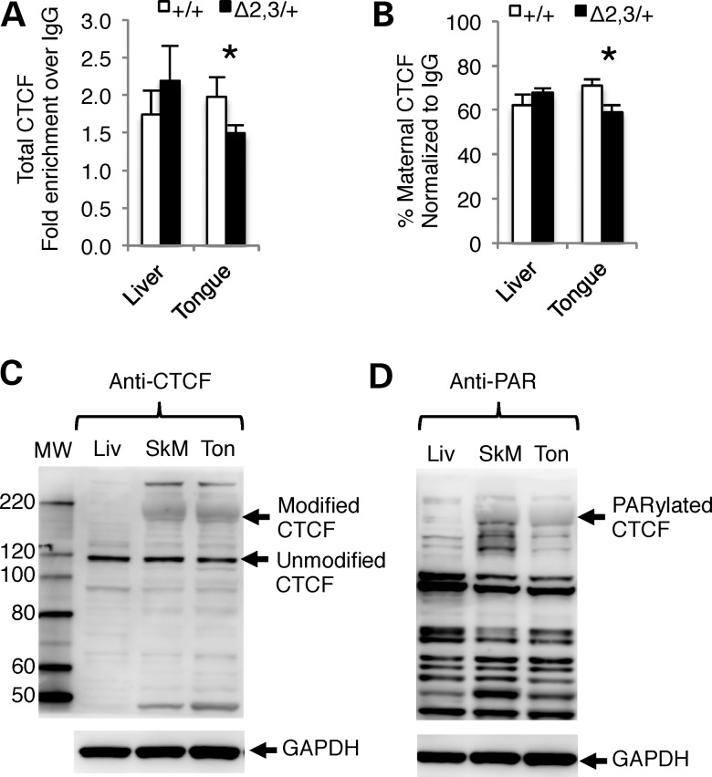
Parent-of-origin-specific expression at imprinted genes is regulated by allele-specific DNA methylation at imprinting control regions (ICRs). This mechanism of gene regulation, where one element controls allelic expression of multiple genes, is not fully understood. Furthermore, the mechanism of gene dysregulation through ICR epimutations, such as loss or gain of DNA methylation, remains a mystery. We have used genetic mouse models to dissect ICR-mediated genetic and epigenetic regulation of imprinted gene expression. The H19/insulin-like growth factor 2 (Igf2) ICR has a multifunctional role including insulation, activation and repression. Microdeletions at the human H19/IGF2 ICR (IC1) are proposed to be responsible for IC1 epimutations associated with imprinting disorders such as Beckwith-Wiedemann syndrome (BWS). Here, we have generated and characterized a mouse model that mimics BWS microdeletions to define the role of the deleted sequence in establishing and maintaining epigenetic marks and imprinted expression at the H19/IGF2 locus. These mice carry a 1.3 kb deletion at the H19/Igf2 ICR [Δ2,3] removing two of four CCCTC-binding factor (CTCF) sites and the intervening sequence, ∼75% of the ICR. Surprisingly, the Δ2,3 deletion does not perturb DNA methylation at the ICR; however, it does disrupt imprinted expression. While repressive functions of the ICR are compromised by the deletion regardless of tissue type, insulator function is only disrupted in tissues of mesodermal origin where a significant amount of CTCF is poly(ADP-ribosyl)ated. These findings suggest that insulator activity of the H19/Igf2 ICR varies by cell type and may depend on cell-specific enhancers as well as posttranslational modifications of the insulator protein CTCF.


Figures

Figure 1.
Figure 1.
Generation of targeted H19ICRΔ2,3 allele. (A) Targeting scheme at the H19/Igf2 locus. Illustrated from top to bottom are the wild-type endogenous locus (H19ICR+), the targeting vector (pH19ICRΔ2,3neo), the correctly targeted allele with the neomycin resistance cassette (H19ICRΔ2,3neo) and targeted allele after neor excision (H19ICRΔ2,3). Restriction sites and their relative positions (in kb) to the H19 TSS are indicated above the endogenous locus. Shaded region between endogenous locus and targeting vector indicates regions of homology. Southern probes (A, B and C) are indicated by horizontal lines below the endogenous locus. Also depicted is the H19/Igf2 ICR (white rectangle), CTCF sites (white triangles), H19 exons (gray rectangles), pBluescriptIIKS sequence (bold line) versus 129/Sv mouse DNA (thin line), neor cassette (polka dot box) and loxP sites (black arrowheads). Sequence deleted by the ICRΔ2,3 mutation is indicated by a cross-hatched region. Line arrows indicate direction of gene expression. (B) Southern blots using mouse tissues confirmed correctly targeted alleles using external probe A and EcoRV digest (i), external probe B and StuI digest (data not shown), and internal probe C and SacI digest (ii) as previously described (36).
Figure 2.
Figure 2.
Neonatal pup and tongue weights in H19ICRΔ2,3/+and H19ICR+/Δ2,3pups compared with wild-type littermates. Consistent trends in pup and tongue weight associated with maternal or paternal inheritance of Δ2,3. Charts show average weight in grams (y-axis) of pups with Δ2,3 deletion (filled bars, maternal transmission—black, paternal transmission—gray) compared with wild-type littermates (open bars). Standard error bars are depicted. Numbers above chart indicate average percent change (%Δ) in weight, positive numbers (increase) and negative numbers (decrease). Letter–number combinations below chart indicate dam (letter) and litter (number), e.g. A1 indicates dam “A” and litter “1”; litters are grouped by pup age but otherwise unordered. Pup age in days post parturition and number of pups analyzed per group (n) is listed in columns below dam. Asterisks indicate significant difference (P < 0.05) as determined by two-tailed t-test. (A) Average pup weight with maternal Δ2,3 inheritance. (B) Average tongue weight with maternal Δ2,3 inheritance. (C) Average pup weight with paternal Δ2,3 inheritance. (D) Average tongue weight with paternal Δ2,3 inheritance.
Figure 3.
Figure 3.
H19ICRΔ2,3 inheritance disrupts H19/Igf2 imprinted expression in neonatal tissues. (A) Illustration of allelic expression at the H19/Igf2 locus. Location of allele-specific assays in Igf2 and H19 is shown as horizontal bars B and C below the locus. (B and C) Allele-specific Igf2 or H19 expression in neonatal tissues as determined by reverse-transcriptase PCR and allele-specific digest and migration in acrylamide gel; maternal (m) and paternal (p) alleles migrate at different sizes due to polymorphic restriction digest sites within the amplicon. Tissue being assayed is listed above gel, indicating lanes containing individual mutant (Δ2,3/+ or +/ Δ2,3) and wild-type (+/+) littermate samples; mesodermal lineages (liver and lung), endodermal lineages (tongue and skeletal muscle). (B) Allele-specific Igf2 expression with maternal inheritance of H19ICRΔ2,3 as compared with wild-type littermates. Percent maternal Igf2 expression is listed below each lane for each respective sample. (C) Allele-specific H19 expression with paternal inheritance of H19ICRΔ2,3 when compared with wild-type littermates. Percent paternal H19 expression is listed below each lane for each respective sample. (D) Total H19 or Igf2 expression (relative to Arppo expression) with maternal H19ICRΔ2,3 inheritance in wild type (+/+, open bars, n = 4) or mutant (Δ2,3/+, filled bars, n = 5), with standard error bars presented. (E) Total H19 or Igf2 expression (relative to Arppo expression) with paternal H19ICRΔ2,3 inheritance in wild type (+/+, open bars, n = 4) or mutant (+/Δ2,3, filled bars, n = 5), with standard error bars presented.
Figure 4.
Figure 4.
Effect of 70% deletion of the H19/Igf2 ICR on DNA methylation states. Percent methylation is depicted across the H19/Igf2 locus with maternal (A) or paternal (B) inheritance of the H19ICRΔ2,3 allele. (A and B) Top: the H19/Igf2 locus is depicted with the following features: Igf2 and H19 genes (open boxes with arrows), differentially methylated regions (DMRs, diamonds), ICR (filled box), CTCF sites (R1 and R2, triangles) and Δ2,3 deletion (hashed box). Total percent methylation (as determined by bisulfite conversion, PCR and pyrosequencing) is shown for Igf2 DMR1, DMR2 and the promoter proximal region in shaded columns for the cell types/tissues indicated in the left column for wild-type (+/+) and mutant samples (Δ2,3). Allele-specific methylation (A, maternal allele and B, paternal allele) at the ICR was determined by bisulfite conversion, PCR, cloning and sequencing. Methylated CpGs (filled circles) and unmethylated CpGs (unfilled circles) are depicted in rows for each clone/copy of DNA and columns indicate individual CpG sites (shaded regions indicate CpGs at CTCF sites). Percent methylation was calculated as number of methylated CpGs over total number of CpGs sequenced and is listed beside clones. Asterisks indicate significant difference (P < 0.05) in DNA methylation levels at the Igf2 DMR1 between wild-type and mutant samples as determined by two-tailed t-test, n = 3–5.
Figure 5.
Figure 5.
Tissue-specific differences in CTCF binding at the mutant H19/Igf2 ICR and poly(ADP-ribosyl)ated CTCF protein. (A and B) CTCF binding to the H19/Igf2 ICR as determined by ChIP and PCR amplification of a 145 bp region overlapping the ICR CTCF site most distal to the H19 promoter. (A) Total CTCF binding and (B) percent maternal CTCF binding. Values are normalized to input and depicted as fold enrichment over IgG. n = 3 l (3–4 dpp). Within each litter, wild-type (+/+, open bars) and mutant (−/+ and +/−, filled bars) tissues were pooled separately. Asterisks indicate significant difference (P < 0.05) as determined by paired t-test. (C and D) Depicts CTCF protein levels in representative samples detected by western blot using GAPDH as loading control and protein marker to determine molecular weight (MW) of bands for identity. (C) Protein levels of CTCF isoforms in neonatal liver (Liv), skeletal muscle (SkM) and tongue (Ton) as determined by western blot probed with anti-CTCF antibody. Arrows indicate modified form of CTCF in SkM and Ton and unmodified CTCF in Liv, SkM and Ton. (D) Poly(ADP-ribosyl)ated proteins in neonatal liver (Liv), skeletal muscle (SkM) and tongue (Ton) as determined by western blot probed with anti- PAR antibody. Arrow indicates poly(ADP-ribosyl)ated band with MW identical to modified form of CTCF in (C).

References

    1. Barlow D.P. Genomic imprinting: a mammalian epigenetic discovery model. Annu. Rev. Genet. 2011;45:379–403. - PubMed
    1. Lee J.T., Bartolomei M.S. X-inactivation, imprinting, and long noncoding RNAs in health and disease. Cell. 2013;152:1308–1323. - PubMed
    1. Ideraabdullah F.Y., Vigneau S., Bartolomei M.S. Genomic imprinting mechanisms in mammals. Mutat. Res. 2008;647:77–85. - PMC - PubMed
    1. De Crescenzo A., Coppola F., Falco P., Bernardo I., Ausanio G., Cerrato F., Falco L., Riccio A. A novel microdeletion in the IGF2/H19 imprinting centre region defines a recurrent mutation mechanism in familial Beckwith-Wiedemann syndrome. Eur. J. Med. Genet. 2011;54:e451–e454. - PMC - PubMed
    1. Beygo J., Citro V., Sparago A., De Crescenzo A., Cerrato F., Heitmann M., Rademacher K., Guala A., Enklaar T., Anichini C., et al. The molecular function and clinical phenotype of partial deletions of the IGF2/H19 imprinting control region depends on the spatial arrangement of the remaining CTCF-binding sites. Hum. Mol. Genet. 2013;22:544–557. - PMC - PubMed

Publication types