. 2015 Jan;1848(1 Pt B):363-8.
doi: 10.1016/j.bbamem.2014.05.025. Epub 2014 Jun 2.

REDOR constraints on the peptidoglycan lattice architecture of Staphylococcus aureus and its FemA mutant

Affiliations

REDOR constraints on the peptidoglycan lattice architecture of Staphylococcus aureus and its FemA mutant

Manmilan Singh et al. Biochim Biophys Acta. 2015 Jan.

Abstract

The peptidoglycan of Gram-positive bacteria consists of glycan chains with attached short peptide stems cross-linked to one another by glycyl bridges. The bridge of Staphylococcus aureus has five glycyl units and that of its FemA mutant has one. These long- and short-bridge cross-links create totally different cell-wall architectures. S. aureus and its FemA mutant grown in the presence of an alanine-racemase inhibitor were labeled with d-[1-¹³C]alanine, l-[3-¹³C]alanine, [2-¹³C]glycine, and l-[5-¹⁹F]lysine to characterize some details of the peptidoglycan tertiary structure. Rotational-echo double-resonance (REDOR) NMR of isolated cell walls was used to measure internuclear distances between ¹³C-labeled alanines and ¹⁹F-labeled lysine incorporated in the peptidoglycan. The alanyl ¹³C labels in the parent strain were preselected for C{F} and C{P} REDOR measurement by their proximity to the glycine label using ¹³C¹³C spin diffusion. The observed ¹³C¹³C and ¹³C³¹P distances are consistent with a tightly packed architecture containing only parallel stems in a repeating structural motif within the peptidoglycan. Dante selection of d-alanine and l-alanine frequencies followed by ¹³C¹³C spin diffusion rules out scrambling of carbon labels. Cell walls of FemA were also labeled by a combination of d-[1-¹³C]alanine and l-[¹⁵N]alanine. Proximity of chains was measured by C{N} and N{C} REDOR distances and asymptotic plateaus, and both were consistent with a mixed-geometry model. Binding of an ¹⁹F-labeled eremomycin analog in the FemA cell wall matches that of binding to the parent-strain cell wall and reveals the proximity of parallel stems in the alternating parallel-perpendicular mixed-geometry model for the FemA peptidoglycan lattice.

Keywords: Alanine racemase; Bacterial cell-walls; Solid-state NMR.


Figures

Figure 1
Figure 1
Location of the labels (red, 13C; green, 19F) of FemA peptidoglycan, and (red, 13C; yellow, 31P) of the repeating unit of wall teichoic acid.
Figure 2
Figure 2
Dante-selected C{F} (left) and C{P} (right) REDOR of intact cell walls of the parent strain of S. aureus grown in media containing D-[1-13C]alanine, L-[3-13C]alanine, [2-13C]glycine, and L-[5-19F]lysine with the alanine racemase inhibitor, alaphosphin. Dante differencing with inversion of the glycyl-carbon peak (42 ppm) preceded REDOR dephasing. Four data blocks were collected resulting in spectra with and without Dante irradiation, each with and without 19F (or 31P) dephasing. The Dante differences (ΔS) are shown at the bottom of the figure and are the reference spectra for REDOR dephasing (ΔΔS) shown above. Spinning sidebands are designated by “ssb.”
Figure 3
Figure 3
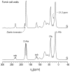
Dante frequency selection for the FemA cell-wall sample. A train of 1-μs 13C radio-frequency pulses separated by 5 μs, with the carrier frequency centered at 178 ppm (top) or 175 ppm (bottom), was followed by z-axis storage for 200 ms. The resulting spectra are designated as “S.”
Figure 4
Figure 4
Dante difference spectra from the FemA cell-wall inversions of Figure 2 showing 13C-13C spin diffusion (for 200 ms) from the carbonyl-carbon label (175 ppm, left; 178 ppm, right) to the glycyl label (near 42 ppm) and the L-alanyl label (15 ppm). ΔS = S0S. Spinning sidebands are designated by “ssb.”
Figure 5
Figure 5
Dante difference spectra from L-alanyl FemA cell-wall inversions showing 13C-13C spin diffusion (for 400 ms) from the methyl-carbon label (17.5 ppm, left; 21.2 ppm, right) to the glycyl label (near 42 ppm) and the D-alanyl label (near 175 ppm). Spinning sidebands are designated by “ssb.”
Figure 6
Figure 6
(Left) C{N} and N{C} REDOR dephasing (ΔS/S0) of 19F-labeled LCTA-1110 complexed to cell walls of the FemA mutant of S. aureus grown on media containing D-[1-13C]alanine and L-[15N]alanine, with the alanine racemase inhibitor, alaphosphin (10 μg/ml), as a function of the dipolar evolution (open circles). The error in the integrated REDOR difference is estimated as the diameter of the open-circle symbols. (Right) C{F} and N{F} REDOR dephasing of the same sample (closed circles). The calculated dephasings for two single-distance components are shown in red and blue, and the combined dephasing curve (25% shorter-distance component and 75% longer-distance component), as a solid line.
Figure 7
Figure 7
(Top left) A cross-section of the proposed peptidoglycan-tertiary structure for the FemA mutant of S. aureus (ref 1). The cross-section consists of sixteen glycan chains in a 4×4 matrix where the glycan backbones (represented by gray circles) are propagating perpendicular to the plane of the paper. The stems and bridges are represented by green and red rectangles, respectively. A cartoon of the glycopeptide LCTA-1110 (bottom right) is shown bound to a D-Ala-D-Ala uncross-linked peptide stem based on the lattice model of ref . Details of the highlighted region of the top-left panel are shown in the light-orange insert (bottom left). REDOR distance measurements (blue numbers) connect labels (red arrows). (Top right) Chemical structure of LCTA-1110.

References

    1. Kim SJ, Singh M, Shasad S, Schaefer J. Cross-link formation and peptidoglycan lattice assembly in the FemA mutant of Staphylococcus aureus. Biochemistry. 2014 doi: 10.1021/bi4016742. - DOI - PMC - PubMed
    1. Kim SJ, Singh M, Preobrazhenskaya M, Schaefer J. Staphylococcus aureus peptidoglycan stem packing by rotational-echo double-resonance NMR spectroscopy. Biochemistry. 2013;52:3651–3659. - PMC - PubMed
    1. Sharif S, Kim SJ, Labischinski H, Schaefer J. Characterization of peptidoglycan in fem-deletion mutants of methicillin-resistant Staphylococcus aureus by solid-state NMR. Biochemistry. 2009;48:3100–3108. - PMC - PubMed
    1. Gullion T, Schaefer J. Rotational-echo double-resonance NMR. J Magn Reson. 1989;81:196–200. - PubMed
    1. Gullion T, Schaefer J. Detection of weak heteronuclear dipolar coupling by rotational-echo double-resonance NMR. Adv Magn Reson. 1989;13:57–83.

Publication types

MeSH terms

LinkOut - more resources